To the Editor,
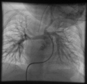
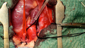
A 9-month-old infant with Down syndrome was in follow-up at our clinic with extensive subarterial ventricular septal defect, stenosis at the origin of both pulmonary vessels, and persistently elevated pulmonary artery pressure. Clinically, she was asymptomatic, with normal weight and height for her age. She was in treatment with digoxin and enalapril. Of note in the physical examination was a difference in saturation between one arm and the other (oxygen saturation by pulse oximetry of 96% in the right arm and 82% in the left arm). Cardiac catheterization was performed to determine the QP/QS ratio and the anatomy of the pulmonary artery branches; a ventricular septal defect was confirmed. There was the additional finding that the left brachiocephalic trunk from which commenced the common left common carotid artery and the left subclavian artery with origin in the pulmonary trunk (Figure 1) formed a right aortic arch. The origin of the right subclavian artery and the right carotid artery were normal. Our patient underwent corrective surgery (Figure 2). The findings described above were confirmed, and the left brachiocephalic trunk was found to be connected to the origin of the left pulmonary artery in the pulmonary trunk via a patent ductus arteriosus. The left brachiocephalic trunk was reimplanted to the aortic arch, the origin of the pulmonary branches was extended, and the defect was closed with a percardial flap. Her postoperative progress was very satisfactory, and she was discharged one week after the operation.

Figure 1. Pulmonary arteriograph in the posteroanterior projection in which the origin of the pulmonary trunk through the ductus arteriosus of the left brachiocephalic trunk is visualized.

Figure 2. Surgical confirmation of the anomalous origin of the brachiocephalic trunk.
The anomalous origin of the left brachiocephalic trunk at the level of the pulmonary trunk is an infrequent vascular malformation. Only 5 cases have been described in the literature, and these always have had the origin in the common carotid artery in the main pulmonary artery,1 but not the brachiocephalic trunk as in our case. Such malformations are associated with anomalies in the aortic arch (essentially in the right aortic arch), with the anomalous origin of one of the subclavian arteries, and with septal defects. In the past, embryogenesis of this malformation has been explained exclusively by anomalies in the regression of the branchial arches,2 but more recently it has been proposed to arise from poor septal formation of the aortic sac due to abnormal migration of cells from the neural crest at the cardiac level.1 Our case could confirm this second theory, given the associated subarterial ventricular septal defect in our patient. The connection of the vessel with anomalous origin is established by means of a patent ductus arteriosus with the trunk of the pulmonary artery at the level of the origin of the left pulmonary artery, as reported in the literature.1, 3.
The different imaging techniques were essential to arrive at an appropriate diagnosis, given the unusual clinical presentation. The outcome of the correction of the malformation was satisfactory.
.
Corresponding author: lopolipillapa@hotmail.com