To the Editor,
Aneurysm of the atrial appendages is an uncommon condition, with few cases described in the literature. These aneurysms are classified into 2 groups: congenital and acquired (mainly secondary to trauma).1, 2 Those affecting the left atrial appendage, which were first described by Semans and Taussing in 1938, usually manifest in the third and fourth decade of life as tachyarrhythmia refractory to pharmacological treatment and thromboembolic phenomena.3 Those that affect the right atrium are rarer and there are few published reports, for the most part in adults.1, 2, 3 It is essential to diagnose atrial appendage aneurysm because of the possible complications mentioned above (arrhythmia and pulmonary and systemic thromboembolism).1 Although sporadic reports have described aneurysm of the right atrial free wall diagnosed in newborns and even prenatally, according to our review, this is the first case of aneurysm of the right atrial appendage described in a neonate. The large size of the aneurysm and the controversial management in this type of patient are highlighted.
A 2-day-old male infant was referred to the cardiology department with a prenatal diagnosis of dilatation of the right atrium (RA), with no other malformations. There were no other prenatal data of interest. The delivery was normal, at term, and there were no incidents. On physical examination, the infant was in good general condition, with good color and peripheral perfusion. On chest auscultation, the heart sounds were rhythmic and clear, and there was good bilateral ventilation. There were no other relevant clinical findings. Chest radiography disclosed considerable cardiomegaly due to RA dilatation, with no other alterations. On electrocardiography, he presented sinus rhythm at 120 bpm, without pre-excitation or other anomalies. Echocardiography in a subcostal view showed a large intrapericardial structure, anterior and to the right of the RA, and communicating with it (Figure 1). The right appendage was very dilated, and was positioned together with the RA, as if it were a third atrium. Color Doppler showed low-velocity flow in the interior. A concomitant, 2-cm permeable foramen ovale with left to right flow was detected. The lesion was confirmed by computed tomography, which depicted a giant aneurysm of the right appendage, measuring 32×25 mm and causing slight compression of the right ventricle (RV) (Figure 2).

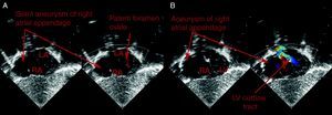
Figure 1. A, Subcostal echocardiographic view. A giant aneurysm of the right appendage is seen, appearing as if it were a third atrium directly communicating with the right atrium. The interatrial septum shows mild flow from the left atrium through the permeable foramen ovale. B, The large size of the aneurysm in relation to the right atrium is evident. Color Doppler shows low-velocity flow in the interior. LA, left atrium; LV, left ventricle; RA, right atrium.

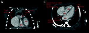
Figure 2. Computed tomography. A, Coronal scan shows the aneurysm slightly compressing the right ventricle. B, Four-chamber view, in which the relationship of the aneurysm with the right chambers is seen. LA, left atrium; LV, left ventricle; RA, right atrium; RV, right ventricle.
Taking into consideration the thrombogenic potential of the lesion, the patient's age, and the electrocardiography findings, antiplatelet therapy and ambulatory monitoring were established, with a plan to have reparative surgery at an older age, if required.
At 1 year 6 months of life, the patient remains asymptomatic, with good morphological and functional development. The lesion has been monitored with periodic ambulatory echocardiography, which has shown no significant changes in size and no internal thrombi. The foramen ovale closed spontaneously, and electrocardiography and Holter recordings remain normal.
Aneurysms of the right appendage are a rare malformation of unknown origin. It is believed that the congenital type, as was seen in our case, is caused by dysplasia of the muscular wall of the atrium.4 The aneurysm may be asymptomatic initially, manifesting mainly in the third and fourth decade of life with symptoms of dyspnea, precordial pain and palpitations. A correct diagnosis is essential because the condition is associated with considerable morbidity, and it should be excluded in cases of arrhythmia refractory to treatment, particularly atrial fibrillation,3, 5 and thromboembolic phenomena.
The diagnosis can be established with fairly non-invasive techniques, such as transthoracic and transesophageal echocardiography,2 computed tomography, and magnetic resonance imaging. Other associated anomalies have been described, such as interatrial communication, persistent left superior vena cava, and anomalous pulmonary drainage, which should also be investigated in these patients.1.
A dilated right atrium can sometimes be mistaken for other, more common conditions, such as Ebstein anomaly.3, 4 The main manifestations of this disease are an abnormal cardiac silhouette and arrhythmic phenomena. The differential diagnosis is can be easily made by evaluating the tricuspid valve insertion.
The natural evolution of these malformations remains uncertain. In patients with aneurysms of the left appendage, even those who are asymptomatic, surgery is recommended because of the high risk of systemic thromboembolism.3 In cases in which the right appendage is affected, there are no other associated defects, and the patient is asymptomatic, management is more controversial. In some studies, a reduction in the risk of atrial arrhythmia, and even resolution of rhythm abnormalities have been demonstrated following surgical excision.4, 5 Oral anticoagulant administration is recommended in patients for whom surgery is not contemplated to reduce the thromboembolic risk.1 In any event, asymptomatic patients should be evaluated individually for treatment.
In our case, an essentially non-aggressive treatment with antiplatelet therapy was decided because of the young age of the patient and the absence of symptoms. The evolution over time will determine whether other, more aggressive treatments, such as surgery are required.
.
Corresponding author: mangelestejero@gmail.com