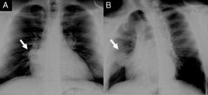
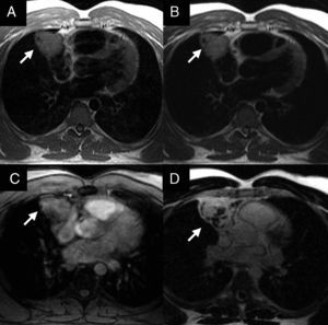

A 34-year-old man was referred for evaluation of a mediastinal mass, detected on routine chest radiography (Figure 1A and 1B). His past medical history, physical examination, and laboratory tests were unremarkable. Cardiac magnetic resonance confirmed a well circumscribed mass in the anterior mediastinum, measuring 6×5×5cm. It started at the level of the pulmonary artery bifurcation and extended laterally to the right atrium, without evidence of infiltration of surrounding tissues. In comparison with the myocardium, the mass was isointense on T1-weighted imaging (Figure 2A) and hyperintense on T2 sequences (Figure 2B), findings compatible with high fluid content. On the other hand, first-pass images showed hyperperfusion within the mass (Figure 2C) and diffuse delayed enhancement (Figure 2D), suggesting a highly vascular component with increased interstitial space. The patient underwent surgical mass resection. Histological specimens showed a central component of large polygonal cells with clear cytoplasm and central nuclei with prominent nucleoli (Figure 3A and 3B). This morphologic finding together with immunohistochemical staining that was positive for placental alkaline phosphatase and negative for alpha-fetoprotein and human chorionic gonadotropin were consistent with thymic seminoma. Germ-cell tumors are the most common cancer in men between the ages of 15 and 35 years. The anterior mediastinum is the most common primary extragonadal site for seminoma. Because of its favorable prognosis, these variants should not be confused with other mediastinal tumors. A cardiac magnetic resonance provides a comprehensive characterization of this tumor, and should be considered when a mediastinal mass is discovered in a young male.

Figure 1.

Figure 2.

Figure 3.
FundingThis study was funded in part by the Sociedad Española de Cardiología and the Instituto de Formación e Investigación “Marqués de Valdecilla” (IFIMAV) in Santander, Spain.
Corresponding author: Javier.Sanz@mssm.edu