To the Editor,
We present the case of a 31-year-old female native of Colombia, with no cardiovascular risk factors or relevant medical history, who was referred to a cardiologist for oppressive chest pain on effort of possible coronary origin and of 5 years of evolution. On physical examination there were no pathological findings of interest. ECG showed a sinus rhythm of 50 bpm, without repolarization alterations; chest X-ray was normal; transthoracic echocardiogram showed a normal sized left ventricle with preserved function, without any changes in general and segmental contractility. A maximum stress test was performed according to the Bruce protocol, which resulted in positive clinical signs and negative electrical signs for ischemic heart disease.
With these findings and given the patient's low risk of ischemic heart disease, a cardiac computed tomography (CT) angiography was requested. This is a non invasive anatomical imaging study which makes it possible to assess the coronary arteries and characterise atherosclerotic plaque1 (it must be kept in mind that it does not provide functional coronary information.) The feasibility and diagnostic accuracy of this study have improved due to widespread use of 64-slice multidetector CT scanners. Current systems provide a clear view of the main coronary arteries and their branches, with a spatial resolution similar to that of conventional angiography.2 The usefulness of this technique is that it provides a maximum negative predictive value (99%-100%) to rule out coronary disease.3 Thus, this technique significantly influences the stratification of selected patients with low or intermediate risk who come to the emergency department with chest pain.2
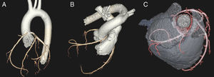
A triple rule-out study was performed after informing patients and obtaining their informed consent for inclusion in an institutional protocol. It was negative for pulmonary thromboembolism and acute aortic syndrome, while non-invasive coronary angiography (64-slice helical CT in prospective acquisition with volume reconstruction performed in sinus rhythm and calcium score 0) showed codominance; normal circumflex artery and left anterior descending artery and its branches; and an anomalous right coronary artery arising from the left sinus of Valsalva (Figure 1A) with decreased calibre due to hypoplasia of the ostium and a sharp angled origin in the aorta. Furthermore, its course ran between the pulmonary artery and the ascending aorta (Figure 1B and C). All these findings indicate a high risk anomalous origin of the right coronary artery.

Figure 1. 64-slice coronary computed tomography (prospective acquisition) with volume reconstruction: an anomalous right coronary artery arising from the left sinus of Valsalva, decreased caliber and interarterial course.
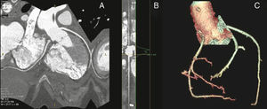
Congenital anomalous origin of coronary arteries is seen in 1% to 1.2% of all coronary angiograms. 0.5% of them show high-risk lesions of the left main trunk or anterior descending branch with their origin in the opposite sinus of Valsalva. Coronary anomalies are the cause of 15% of sudden deaths in athletes. In 80% of the autopsies of athletes after sudden death with coronary arteries of anomalous origin, the affected coronary artery runs between the aorta and the pulmonary artery. Currently, the anatomical description of a coronary artery that passes between the aorta and pulmonary artery in a young person (under 50) is a major risk factor for an adverse event, with or without symptoms.4In this instance, after diagnostic confirmation by invasive coronary angiography, we opted for surgical revascularization by coronary reimplantation (AHA/ACC3 class I indication). It is worth noting that we were able to confirm the anomalous origin and interarterial course of the coronary artery, and furthermore, we found a 2cm intramural course beginning at the mid-surface of the right sinus. We reimplanted the right coronary artery in the non-coronary sinus, without complications and with a good outcome (Figure 2A-C).

Figure 2. Right coronary artery reimplanted in the non-coronary sinus, without residual stenosis.
.
Corresponding author: M.CRISTINA.GOENAVIVES@osakidetza.net