We report the case of a 57-year-old man with a 3-month history of intermittent pyrexia. He received irregular antibacterial therapy and thrombolysis in a local hospital due to occlusion of the right distal popliteal artery. His medical history included multiple fractures, allergic purpura, and hypertension. His medications included prednisone (30mg orally per day), metoprolol, and amlodipine.
On physical examination, breath sounds were clear and a 3/6 pansystolic murmur was auscultated at the right sternal border. Swelling of the right leg and gangrene at the fifth toe were found. Abdominal palpation revealed mild splenomegaly. The following abnormal laboratory results were identified: white cell count, 3.20 × 10⁹/L; platelet count, 30 × 109/L; hemoglobin, 8.80g/dL; albumin, 2.99g/dL; erythrocyte sedimentation rate, 42mm/h; C-reactive protein, 13.30mg/L; and ferritin, 674μg/L.
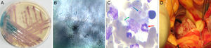
Three blood cultures were positive and a gram stain showed budding yeast cells. The isolate, after being subcultured on CHROMagar (Becton Dickinson, Paris, France), showed membranous colonies that changed color from pink to blue within 48hours (Figure A). On corn meal agar (Becton Dickinson), pseudohyphae and blastoconidia were seen 24hours later (Figure B). The yeasts were identified as Kodamaea ohmeri (K. ohmeri). Drug sensitivity testing showed that this strain was susceptible to voriconazole, fluconazole, itraconazole, and amphotericin B.
, multiple red blood cells (green arrow), and platelets (blue arrow) (Giemsa stain; magnification, ×1000). D: Intraoperative view shows a large fragile and loose vegetation on the aortic valve that almost occludes the orifice.")
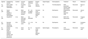
A: Color change of K. ohmeri cultured on CHROMagar Candida medium. B: Subcultured on corn meal agar, the yeast shows pseudohyphae and blastoconidia. C: Bone marrow smear shows macrophage phagocytosis of a band neutrophil (red arrow), multiple red blood cells (green arrow), and platelets (blue arrow) (Giemsa stain; magnification, ×1000). D: Intraoperative view shows a large fragile and loose vegetation on the aortic valve that almost occludes the orifice.
Bone marrow aspiration, performed due to the cytopenia, showed phagocytosis of hematopoietic cells by activated macrophages (Figure C). Thoracoabdominal computed tomography revealed splenomegaly and mild bilateral pleural effusion. Transthoracic echocardiography showed a large vegetation (30mm × 12mm) on the aortic valve with mild regurgitation and stenosis.
On hospital day 5, the patient developed persistent pyrexia with a temperature of 39°C despite antifungal therapy with intravenous voriconazole. Urgent surgery was performed and a large fragile and loose vegetation was found on the aortic valve that almost occluded the orifice (Figure D). The aortic valve was replaced with a 21-mm mechanical prosthesis (St. Jude Medical, St. Paul, MN, United States). Continued blood loss of more than 200mL/h occurred in the postoperative period. The coagulation profile showed hypocoagulability with fibrinogen of less than 1.5g/L and a platelet count of 20 × 109/L. Hemophagocytic lymphohistiocytosis (HLH) was diagnosed and the patient was started on intravenous methylprednisolone 500mg/d and immunoglobulin 0.5g/kg/d for 3 days and transfusion of packed red blood cells, plasma, platelets, cryoprecipitate, and fibrinogen. The treatment effectively stopped the bleeding. The patient eventually recovered well and was discharged.
HLH is rare and has a mortality of 41%. It is characterized by defective cytotoxic cell control of an initial immune response that progresses to uncontrolled macrophage activity and hypercytokinemia. This activation produces an exaggerated inflammatory response and hypersecretion of cytokines in a so-called cytokine storm.1
According to the HLH-2004 diagnostic guidelines, our case fulfilled 6 of the 8 criteria, namely, fever, splenomegaly, cytopenia, fibrinogen ≤ 1.5g/L, hemophagocytosis in the bone marrow, and ferritin ≥ 500μg/L. After reviewing 2197 adult HLH cases, Ramos-Casals et al.1 determined that viral infection was the most common cause of HLH (34.68%); fungus (1.68%) was a less frequent trigger. K. ohmeri is a rare fungal pathogen in humans. There is no published report of secondary HLH related to K. ohmeri infection and we believe it to be a new trigger of HLH.
K. ohmeri is commonly used in the food industry for its fermentation ability. The first case of human infection was reported in 1998, and the pathogen, isolated from pleural fluid, was considered a contaminant.2K. ohmeri endocarditis has thus far only been reported in 4 patients.3–6 The characteristics of all 5 cases (including the present patient) are described in the Table. The patients were all male with predisposing factors. The mitral valve was the most commonly involved valve. The present patient is the first reported case with aortic valve involvement. Vegetations were larger than 10mm in 4 of the 5 patients. This feature is associated with fungal endocarditis and favors the occurrence of embolic events. A combination of valve replacement and an antifungal drug was used to treat K. ohmeri endocarditis in 4 of the 5 patients. One patient, treated by antifungal therapy alone, died before planned surgery could be performed; consequently, we believe that surgery should be urgently performed to treat K. ohmeri endocarditis. Compared with the previously reported cases, the present patient had a longer duration that induced more severe multiorgan injuries, which may be the potential cause of the associated HLH.
Summary of the Characteristics of the Patients With K. ohmeri Endocarditis
| Age/sex | Predisposing factor(s) | Duration | Involved valves | Vegetation dimension | Embolic event | Organomegaly | Hemocytopenia | Treatment | Result | Reference |
|---|---|---|---|---|---|---|---|---|---|---|
| 76 y/M | MVR+CABG, pacemaker implantation | 1 d | Prosthetic mitral valve | 5-cm length | Splenic infarction | No | Thrombocytopenia | MVR, pacemaker removal, AmB lipid complex | Recovered | Reina et al.3 |
| 42 y/M | Heroin addict, hepatitis C, peripheral vasculopathy | 2 wk | Mitral valve with rheumatic disease | 20mm in largest diameter | Occlusion of the distal left popliteal artery | No | Erythropenia | Embolectomy, MVR, liposomal AmB | Recovered | João et al.4 |
| 43 y/M | Hepatitis B | 2 mo | Mitral valve with rheumatic disease | Not described | No | No | Erythropenia | MVR+TVP, itraconazole | Recovered | Yanghua et al.5 |
| 40 d/M | Neonatal seizures, necrotizing enterocolitis | 20 d | Tricuspid valve | 11.1 × 6.4 mm | No | No | Erythropenia | AmB | Death | Sundaram et al.6 |
| 57 y/M | Multiple trauma, broad-spectrum antibiotic therapy, long-term use of prednisone | 3 mo | Aortic valve | 30 × 12 mm | Occlusion of the distal right popliteal artery | Splenomegaly | Pancytopenia | AVR, voriconazole | Recovered | Present case |
AmB, amphotericin B; AVR, aortic valve replacement; CABG, coronary artery bypass grafting; M, male; MVR, mitral valve replacement; TVP, tricuspid valve plasty.
In the present case, K. ohmeri emerged as a rare infectious fungus and was accompanied by a life-threatening HLH, which undoubtedly complicated endocarditis diagnosis and treatment. We believe that multidisciplinary collaboration can favor early recognition and diagnosis and will eventually result in a specific therapy.
This work was supported by the Priority Academic Program Development of Jiangsu Higher Education Institutions [JX10231801].