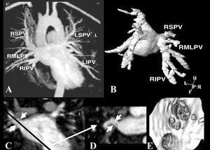
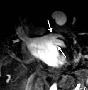
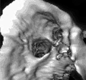
Accurate knowledge of the anatomy of the pulmonary veins is important in clinical electrophysiology. In order to evaluate the usefulness of magnetic resonance angiography for this purpose, we studied 17 unselected patients. All the pulmonary veins were visualized in each individual. The diameters of the ostia ranged between 9 mm and 22 mm. The cross-section of the ostium was elliptical in 35% of cases. In 14 patients (82%), the 4 veins each had independent drainage. In 2 patients (12%), there was an additional intermediate right vein and, in 1 patient (6%), both left veins had a common ostium. In 74% of patients, the right pulmonary veins had a short common trunk with early branching. This pattern was seen in only 10% of left veins. Magnetic resonance angiography using a contrast medium is an excellent technique for studying the anatomy of the pulmonary veins and for identifying variants. The resulting information is potentially useful for electrophysiologists.
Keywords
Magnetic resonance imaging
Pulmonary veins
Catheter ablation
Atrial fibrillation
Este artículo solo puede leerse en
pdf
Bibliography
[1]
Ho SY, Cabrera JA, Tran VH, Farre J, Anderson RH, Sánchez-Quintana D..
Architecture of the pulmonary veins: relevance to radiofrequency ablation..
Heart, (2001), 86 pp. 265-70
[2]
Vonken EA, Velthuis BK, Wittkampf FH, Rensing BJ, Derksen R, Cramer MM..
Contrast-enhanced MRA and 3D visualization of pulmonary venous anatomy to assist radiofrequency catheter ablation..
J Cardiovasc Magn Res, (2003), 5 pp. 545-51
[3]
Dill T, Neumann T, Eikinci O, Breidenbach C, John A, Erdogan A, et al..
Pulmonary vein diameter reduction after radiofrequency catheter ablation for paroxysmal atrial fibrillation evaluated by contrast-enhanced three-dimensional magnetic resonance imaging..
Circulation, (2003), 107 pp. 845-50
[4]
Anatomía humana. 8.ª ed. Barcelona: Salvat Editores, S.A.; 1981. p. 408-10.
[5]
Anatomía Humana. vol. III. 4.ª ed. Barcelona: Editorial Científico-Médica; 1972. p. 225-30.
[6]
Gray's anatomy of the human body. 13th ed. Philadelphia: Lea & Febiger; 1985. p. 794-7.
[7]
Wittkampf FH, Vonken EJ, Derksen R, Loh P, Velthuis B, Wever EF, et al..
Pulmonary vein ostium geometry: analysis by magnetic resonance angiography..
Circulation, (2003), 107 pp. 21-3
[8]
Tsao HM, Wu MH, Yu WC, Tai CT, Lin YK, Hsieh MH, et al..
Role of right middle pulmonary vein in patients with parxysmal atrial fibrillation..
J Cardiovasc Electrophysiol, (2001), 12 pp. 1353-7
[9]
Takase B, Nagata M, Matsui T, Kihara T, Kameyama A, Hamabe A, et al..
Pulmonary vein dimensions with paroxysmal atrial fibrillation using magnetic resonance angiography..
Jpn Heart J, (2004), 45 pp. 81-92
[10]
Tsao HM, Yu WC, Cheng HC, Wu MH, Tai CT, Lin WS, et al..
Pulmonary vein dilation in patients with atrial fibrillation: detection by magnetic resonance imaging..
J Cardiovasc Electrophysiol, (2001), 12 pp. 1333-4