Assessment of the coronary anatomy in patients with Kawasaki disease can be problematic when there is no clear indication for an invasive study. We present a clinical case in which multislice computed tomography (CT) coronary angiography was essential for the diagnosis.
A 16-year-old patient with a prior history of Kawasaki disease underwent echocardiography to investigate suspected coronary aneurysms, with inconclusive results. The patient had no other relevant medical data and was asymptomatic.
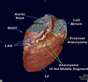
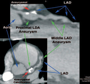
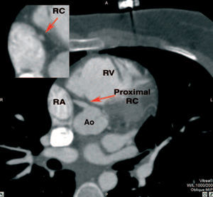
The multidetector CT study showed aneurysms in the proximal (saccular, 9x15 mm) and middle (5x5 mm) left anterior descending artery (Figure 1). Reconstruction of the lumen by multiplanar (Figure 2, upper image) and maximum intensity projection (Figure 2, lower image) reconstruction techniques ruled out the presence of stenotic segments. The circumflex artery had an independent origin and no lesions (not shown). A fusiform aneurysm was detected in the right coronary artery, affecting the proximal 15 mm (Figure 3, compare the diameter of the vessel in the proximal area with that of the healthy portion in the detailed view). Isotope perfusion imaging was performed with negative results, and aspirin therapy was started at 100 mg/day.

Figura 1.

Figura 2.

Figura 3.
Multidetector CT is an imaging tool that is gradually being incorporated into clinical practice, as it allows non-invasive assessment of the coronary anatomy, making it a useful option for the evaluation and follow-up of patients with sequelae of Kawasaki disease.