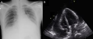
We present the case of a 31-year-old man with no medical history of interest, who presented to the emergency room of a regional hospital with a 1-week history of asthenia and general malaise, associated with diffuse abdominal discomfort, dry cough, and predominantly nocturnal sweating. Physical examination detected hypophonesis in both lung bases and an increase in jugular venous pressure. Laboratory analyses showed bicytopenia (leukocytes, 3800/μL; hemoglobin, 10.7 g/dL) and C-reactive protein concentration at 5.90 mg/dL (upper normal limit, 0.5 mg/dL). Chest radiography depicted cardiomegaly and bilateral pleural effusion. As part of the diagnostic study, transthoracic echocardiography was requested. The examination showed severe pericardial effusion with echocardiographic evidence of tamponade (Figure), which prompted his transferal to our center.

The patient was admitted to the cardiac critical care unit, where pericardiocentesis was successfully carried out, yielding 1350 mL of serohematic fluid. On biochemical analysis, the fluid was identified as an exudate (proteins, 3.1 g/dL; fluid/plasma protein ratio, 0.57; lactate dehydrogenase, 1295 U/L), with an adenosine deaminase level of 26.4 U/L and abundant red blood cells. Cytology showed atypical lymphocytes with an immunoblastic appearance, prompting a request for flow cytometry study. Cytometry identified 54% of hematopoietic cells (CD45), which were large (elevated SSC) and negative for B-cell antigens (CD19) and T-cell antigens (CD3). They were, however, positive for activation antigens, such as HLA-DR and CD30, and for plasma cell markers, such as CD38 and CD138. This immunophenotype in serous effusion is diagnostic of primary effusion lymphoma (formerly known as body cavity lymphoma), a type of large B-cell lymphoma with plasmablastic differentiation associated with human herpes virus 8 (HHV-8) and typically affecting immunosuppressed patients. Serological analysis was then requested, which was positive for human immunodeficiency virus (HIV-1) (chemiluminescence with Western blot confirmation) and HHV-8 infection (immunoglobulin G titer, 1/160, and positive polymerase chain reaction testing). In the assessment of disease extension, there was no evidence of lymphadenopathies or hepatosplenomegaly, and bone marrow biopsy yielded normal results. Pericardial fluid culture was negative.
Based on these findings, a diagnosis of primary effusion lymphoma associated with HIV/AIDS (Category C) was established. CD4 cell count at the time of diagnosis was 104 cells/μL and viral load was 103 000 copies/mL. Antiretroviral treatment (ART) was started with emtricitabine, tenofovir, and raltegravir, and polychemotherapy was subsequently added (DA-EPOCH regimen).
In 2015 in Spain, 3428 new cases of HIV infection were notified, representing a rate of 7.39 new HIV diagnoses per 100 000 population per year. Almost half of these cases were late diagnoses, when CD4 counts were < 350 cells/μL.1 Since the introduction of ART, the prognosis of HIV patients has radically changed, with a considerable increase in survival. In addition, this treatment has led to a decrease in associated pericardial, myocardial, and valvular heart diseases. In the pre-ART era, pericardial disease was the most common cardiac manifestation in HIV patients,2,3 and the estimated prevalence of pericardial effusion was 11%.4 Although these effusions are mainly idiopathic, some may be related to opportunistic infections or various types of cancer, such as lymphoma or Kaposi sarcoma. For this reason, HIV infection should be included in the differential diagnosis of pericardial effusion of uncertain etiology.5
Primary effusion lymphoma is an uncommon (4% of HIV-associated lymphomas) and very aggressive form of non-Hodgkin, diffuse, large B-cell lymphoma that develops exclusively in severely immunosuppressed individuals, generally patients with HIV and very low CD4 lymphocyte counts. Affected patients characteristically have B symptoms (fever, weight loss, night sweats) associated with lymphomatous pleural, pericardial, or peritoneal effusion, without tumor masses, lymphadenopathies, organomegaly, or bone marrow infiltration. The causal agent is HHV-8, the same virus that causes Kaposi sarcoma. This type of lymphoma has a very poor prognosis, with an overall survival of < 6 months. However, prognosis can be improved by the use of ART.6 The patient reported underwent 6 cycles of chemotherapy, and at the time of writing, is in complete remission with an undetectable viral load, although he has still not achieved immune reconstitution.
Because of the aggressiveness and unfavorable prognosis of this type of lymphoma, it could be advisable to include it in the initial clinical suspicion whenever HIV-infected patients develop pericardial effusion of uncertain etiology. Flow cytometry is an important diagnostic technique for these cases, as it allows fast identification of lymphomatous cells suspended in the infiltrated fluid. An early diagnosis would enable initiation of targeted treatment that could improve survival and quality of life in these patients
.