Keywords
INTRODUCTION
Persistent patency of the foramen ovale into adulthood has clinical implications.1 Echographic detection of patent foramen ovale (PFO) is based on evidence of the early passage of microbubbles to the left chambers of the heart or the systemic arteries after intravenous injection of a first-generation echo contrast, usually agitated saline solution.2,3 It has been shown that femoral injection increases the sensitivity of the technique, when compared to antecubital injection.4,5 This difference has been assumed to be due to the orientation of the right atrial-inferior vena cava junction facing the fossa ovalis, with contribution of the Eustachian valve.2-5 However, improved right atrial opacification after contrast injection into a central vein has not been specifically investigated. This study compares the results of injections of agitated saline solution into the femoral vein and the antecubital vein on intracardiac echocardiography (ICE) of the atria.
METHODS
The study included patients scheduled for electrophysiological study (EPS) requiring transseptal puncture. All patients underwent transthoracic echocardiography (TTE) using second harmonic imaging and transesophageal echocardiography (TEE) in the 48 h before EPS, including a study with agitated saline for detection of patent foramen ovale (PFO).6 The agitated saline study by TTE and TEE consisted of a baseline injection and another with the Valsalva maneuver (VM), for which patients had been previously instructed. The release phase of the VM was made to coincide with the time point at which contrast clearly filled the right atrium. An intracardiac echocardiography (ICE) was also done during the EPS to assist with transseptal puncture.7 All patients received intravenous sedation with continuous propofol perfusion (effect-adjusted dose around 8 mg/kg/h). A radial imaging catheter with mechanical rotation was used for intracardiac echocardiography (Ultra ICETM 9F/9MHz, Boston Scientific Corp; USA) and an intravascular ultrasound console (Galaxy2 TM IVUS Imaging System, Boston Scientific Corp; USA). The catheter was advanced to the right atrium and positioned in the region of the fossa ovalis, and image gains were adjusted to obtain slight background noise. A right antecubital venous line was cannulated with an 18 G catheter, and the right femoral vein with a 4 Fr catheter. Using two 10-mL syringes connected to a three-way valve, contrast was obtained by brisk passage of 9.5 mL of normal saline and 0.5 mL of air between the 2 syringes. Ten mL of saline contrast were subsequently injected into the antecubital and femoral vein, ensuring that the contrast of the previous injection was not visible. In the ICE study, only 1 injection was performed per each route to simplify the procedure. The VM was carried out in each of these injections. The ICE images were recorded in DICOM and super-VHS format for subsequent interpretation. Two cardiologists with experience in contrast and intracardiac echocardiography scored by consensus the degree of enhancement in the right atrium from 0 to 4 (0=absence of echo contrast, 4=dense, homogeneous opacification of the right atrium). The images of contrast injections from the same patient were never shown consecutively and the observer was unaware of the injection site used for each image. The Wilcoxon t test was used to compare the degree of opacification.
RESULTS
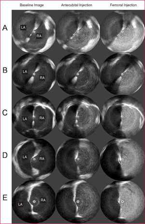
The characteristics of the 21 patients included in the study are shown in Table. No patient had structural heart disease and no complications were caused by the study procedures. Contrast injection via the antecubital route achieved a medium degree of intensity of 1.62 (0.67), whereas the medium opacification achieved by femoral route was 3.76 (0.44). This difference was statistically significant (P<.001). The Figure shows the type of image obtained. Foramen ovale patency could not be demonstrated by intracardiac echocardiography in any patient, despite high-grade imaging and prolonged recording of images after the injection. Previously, PFO was detected in 14% of these patients by TEE and in 33% by TTE (P=NS), by early passage of bubbles to the left atrium.


Figure. Intracardiac ultrasound images. Rows A-E: representative cases. First column: baseline image of intracardiac echocardiography. Second column: echo contrast of right atrium after antecubital injection of agitated saline. Third column: echo contrast of right atrium after femoral injection. Note that opacification is more intense after femoral versus antecubital injection. LA indicates left atrium; RA, right atrium.
DISCUSSION
Two studies4,5 have shown that contrast injection by the femoral vein has greater sensitivity for ultrasound detection of PFO than antecubital injection. This superiority has been attributed to the unique position of the right atrial-inferior vena cava junction, where flow is aimed toward the foramen ovale region.2-5 In these series, the degree of right atrial opacification obtained with each injection type was not specifically monitored. Good, dense contrast in the right atrium could improve PFO detection3 and would obtain a greater demonstrable degree of passage of bubbles into the left heart after a considerable increase of the right atrial pressure.
The present study shows that injection of the same volume of contrast into the femoral vein produces dense, homogeneous opacification of the right atrium, significantly greater than that obtained with injection into the antecubital vein. This fact could have acted as a confounding factor in studies comparing antecubital and femoral injections of agitated saline for the diagnosis of PFO.4,5 Future studies with contrast echocardiography should monitor the degree of right opacification obtained with each injection type.
Patent foramen ovale was not shown in any patient with the use of contrast ICE, even though images with excellent resolution of both atria and the fossa ovalis region were obtained and the patient was asked to perform the VM. There are no previous studies on PFO using contrast ICE; however, given the quality of the images, we doubt that the sensitivity of this ultrasound method is absolutely null, despite our results. All patients had previously performed the VM during TEE and TTE, and some had shown early evidence of shunting. The absence of bubble passage into the left atrium with ICE could be attributed to ineffective performance of the VM during ICE. This observation is consistent with the trend to a lower sensitivity of TEE compared with TTE using second harmonic imaging for PFO detection observed in our study, and is consistent with findings in the literature.6 Although not an explicit part of the protocol, patients at our hospital were usually sedated with midazolam at the operator´s discretion for TEE. In addition to the effect of sedation on the low effectiveness of VM, the possibility that patients with esophageal intubation or central venous lines perform the VM less vigorously should be considered.
Factors other than the injection site could have contributed to the differences observed between the administration routes. Quantitation of the degree of left atrial opacification was blinded for the source of each injection, although the echocardiography specialists were able to identify the outflow of contrast and infer the injection site. However, the differences obtained would outweigh the importance of the necessarily inadequate blinding for the injection site. The femoral injection was done using the same volume of contrast as the antecubital injection, even though the gauge of the femoral catheter would have allowed higher flow. Studies that have previously compared the 2 injections did not control catheter size, and one of them used the femoral vein access for hemodynamic studies of the right side.4,5 Although the expected risk of complications is low, it was not considered ethical to cannulate the femoral vein with an 18 G catheter unless there was an additional clinical purpose. In order to precisely assess the importance of flow direction on the difference between the antecubital and the femoral vein injections of saline contrast for PFO detection, future studies should quantitatively measure opacification of the right atrium.
In conclusion, femoral vein injection of contrast leads to significantly greater density of right atrial opacification than antecubital injection. This should be taken into account in studies that compare antecubital and femoral vein injections of agitated saline for echocardiographic detection of PFO.
Correspondence: Dr. D.R. Saura Espín.
Puerta de Orihuela, 3 bis; 7.o D. 30003 Murcia. España.
E-mail: danielsaura@secardiologia.es
Received October 25, 2006.
Accepted for publication March 1, 2007.