Keywords
INTRODUCTION
Coronary angiography is a reliable diagnostic procedure and its risk of major complications (acute heart attack, cerebrovascular accident) is less than 0.5%, with a mortality rate of less than 0.2%.1
Cortical blindness following the use of contrast agents is uncommon. It was initially described in neurological procedures in 19702; however, it has rarely been seen following cardiac catheterisation. In this article we have outlined our experience of 3 cases following 22 300 coronary angiographies carried out since 1995.
CLINICAL CASES
Case 1
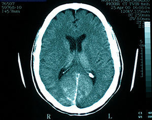
Patient 61 years of age, no allergies, hypercholesterolemic, and ex-smoker. Previous coronary angiography carried out with no complications. Patient underwent triple aortocoronary bypass 1 year earlier, a left internal mammary artery (IMA) was implanted to the anterior descendent (AD) and a graft of the right sequential IMA at a first diagonal (D1) and second obtuse marginal (OM2). Hospitalised for atypical coronary angiography symptoms, carried out via the right femoral vein; disorder detected in 2 of the vessels with occlusion of the AD, 90% D1 stenosis and 75% in the proximal circumflex (Cx). The grafts were permeable and undamaged. 300mL of non-ionic contrast agent (Omnipaque 350 mg/mL) was used and 5000 U of unfractionated heparin. During catheterisation of the left IMA, the patient suffered transient deterioration in the level of consciousness and posterior complete bilateral visual deficit. Cerebral arteriography was carried out with no pathological findings. Over the following hours the patient suffered from cephalalgia and progressive recuperation of vision after 12 hours. Renal functioning was normal pre and post catheterization. The cerebral computerised tomography (CT scan) at 48 hours showed persistent enhancement in the occipital region (Figure 1). Three days later the patient was discharged having completely regained the vision. Four years later the patient underwent another catheterisation with no complications.

Figure 1. Simple CT scan carried out 48 hours after the procedure in case 1: persistent enhancement of the occipital region can be seen.
Case 2
Patient 75 years of age, hypertensive, hypercholesterolemic, and smoker. Admitted for non-Q AMI. Cardiac catheterisation via the right radial was carried out, which showed evidence of coronary disease of 2 vessels with stenosis of 99% of the middle Cx vessel and OM2, as well as stenosis of 75% of the middle and distal right coronary artery (RCA). Following diagnosis, the patient had an episode of temporal-spatial disorientation which lasted for some minutes and the only neurological deficit was the complete lack of vision, so the procedure was suspended. During the test, there were used 170mL of non-ionic contrast agent (Omnipaque 350 mg/mL), 5000 U of unfractionated heparin and spasmolytic cocktail (2.5 mg of verapamil and 200 µg of intraarterial nitroglycerine). The neurological assessment was otherwise normal. Renal functioning was normal both before and after the catheterisation. After 10 hours, progressive recuperation of visual acuity was recorded and no significant alterations were noted in the CT scan carried out after 12 hours. Recovery was complete after 48 hours. One week later another catheterisation was carried out and the described damages were treated without complications.
Case 3
Patient 78 years of age, hypertensive, and dyslipemic. Nine years earlier the patient underwent quadruple aortocoronary bypass with a terminolateral graft of the left IMA to middle AD, and 3 saphenous grafts to D1, first marginal obtuse coronary artery, and posterior descendent. In the last few years, the patient was diagnosed with nephrotic syndrome of undetermined etiology. Currently suffering from chronic renal failure (creatinine clearance, 22 mL/min; basal creatinine serum, 3 mg/dL), pending dialysis. Admitted for spontaneous angina and coronary angiography carried out via the left radial which detected 90% stenosis in the left IMA graft.
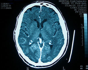
Patient was treated with percutaneous transluminal coronary angioplasty (PTCA) and a stent implantation. We used 624 mL of non-ionic contrast agent (Omnipaque 350 mg/mL, as were 7000 U of unfractionated heparin and spasmolytic cocktail. There were no complications during the test. Bilateral loss of vision occurred after 6 hours. The rest of the neurological examination was completely normal. In the CT scan carried out 2 hours after the onset of symptoms, penetration of the contrast agent was experienced in the arterial tree and the intracranial dural venous sinuses, with persistent enhancement of the occipital region (Figure 2). After 24 hours, an ophthalmologic examination revealed subcapsular posterior cataracts and grade 1 diabetic retinopathy; the complete lack of vision persisted although the patient denied it. Due to worsening of renal function, conventional dialysis was initiated.

Figure 2. Simple CT scan carried out 2 hours after the procedure in case 3: this shows the impregnation of contrast agent in the arterial tree and dural venous and intracranial sinuses with enhancement in the occipital region.
Over the following days visual acuity was recuperated and the patient was discharged after 10 days having almost completely recuperated visual acuity to the point prior to the procedure
DISCUSSION
Cortical blindness is characterised by a loss of visual perception whilst ocular motility and pupillary response are preserved intact. It has been described as a rare complication of AMI occurring in 0.31% of cerebral and vertebral angiographies.3 The estimated incidence in coronary angiography is of 0.05%.4
Its physiopathology is unknown and various mechanisms have been proposed such as the direct toxicity of the contrast agent,5 microembolisms,6 hypoxia, or cerebral oedema. The use of hyperosmolar solutions increases the permeability of the blood-brain barrier which is especially thin in the occipital region, therefore facilitating the way for the contrast agent into the area of the visual cortex due to low localisation during the procedure.7 The least sympathetic innervation of the vertebral territory in comparison with the carotid seems to favour this phenomenon.8 The susceptibility of the blood-brain barrier to permeability is higher in patients with chronic hypertension.9 Renal failure and the quantity and hyperosmolality of the contrast agent have also been suggested as favouring this complication. The fact the majority of cases described have IMA aortocoronary bypass can be explained by the injection of the contrast agent via the vertebral artery during catheterisation of the IMA ostium and the need for a higher amount of the agent in these patients.
The cases presented here coincide with the few that have been published.
Symptoms usually start during the procedure or in the following 12 hours, with varying symptoms which can include cephalalgia, disorientation, nausea, vomiting, or decrease in the level of consciousness, accompanied by the sudden onset of a complete loss of vision. On occasions, as can be seen in our third case, the patient can deny the loss of vision although it is evident when examined. This symptom is known as Anton's syndrome. The lack of vision is progressively recuperated between 6 and 48 hours following the procedure and it has not been demonstrated that the use of diuretics, corticoids, antihistamines, or dextrans accelerates recovery.10 Via a CT scan, an area of increased uptake of the agent can be seen in the occipital region of 2 of the patients, which was resolved with subsequent controls; the radiological descriptions of the abnormal CT scans in our cases coincide with the majority of published cases.
Hinchey et al11 described reversible posterior leukoencephalopathy syndrome (RPLS), consistent with a similar neurological case as the one described, but in a different clinical context (hypertensive individuals with chronic renal insufficiency or undergoing treatment with immunosupressors) and not subjected to examinations with a contrast agent. It is not clear if both are related, and differences are noted in their development, with a slower recovery time in patients affected by RPLS.12
It is a rare disorder, the frequency of which will probably increase over the following years as a consequence of the spreading of this type of examinations. It is of utmost importance to recognise that its transient course does not contraindicate previous explorations, as no recurrences in a second procedure with a contrast agent have been recorded. Although its etiology and treatment continue a mystery, it does seem reasonable to maximise precautions in the catheterisation of the IMA in patients with myocardial revascularisation via this vessel, as well as the use of low osmolality agents in reduced amounts.
Correspondence: Dr. J. García de Lara.
Unidad de Hemodinámica. Hospital Virgen de la Arrixaca.
Ctra. de Madrid, s/n. 30120 Murcia. España.
E-mail: garciadelara@secardiologia.es
Received February 19, 2007.
Accepted for publication June 29, 2007.