Aortic mural thrombus is an uncommon condition with a prevalence of around 1% of all systemic embolisms.1 The development of a thrombus in an aorta with no predisposing lesions is even less frequent, and the treatment of these cases is controversial.2
We present the case of a 45-year-old male smoker with no other cardiovascular risk factors. He had a history of stroke causing an ischemic lesion some years before his current admission, at which time the cardiologic examination was normal. The patient was admitted to our center with diffuse abdominal pain that was more pronounced in the right iliac fossa. He underwent abdominal ultrasound, which yielded normal findings, and abdominal computed tomography (CT), which showed multiple hypodense areas in both kidneys, consistent with renal infarctions.
Based on his history of stroke and the current clinical picture, urgent echocardiography was requested to rule out an intracardiac origin of the embolisms. The echocardiogram showed a nondilated, functionally normal left ventricle, and valvular structures with no anomalies.
Transesophageal echocardiography (TEE) was then carried out, which showed normal heart chambers and no relevant findings. On evaluation of the aorta, a mobile, intra-aortic thrombus, 4cm in length, was observed (video in the supplementary material) on a vessel wall that showed no evidence of a lesion.
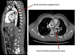
Urgent chest CT study then disclosed a mobile thrombus anchored to the anterior wall of the proximal descending thoracic aorta, posterior to the aortic arch. An atherosclerotic plaque could not be ruled out as the etiologic mechanism (Figure). The angiology and vascular surgery department was consulted, which recommended anticoagulation and posterior re-evaluation.

During hospitalization, the patient had a new onset of diffuse abdominal pain and general deterioration of his clinical status, which prompted a new thoracoabdominal CT study. A portion of the thrombus was seen to have migrated toward the superior mesenteric artery, producing acute intestinal ischemia.
The patient underwent urgent resection of the affected portion of the small intestine, with a side-to-side, jejunojejunal anastomosis. Subsequently, a 26×150-mm Dacron endoprosthesis was implanted. The patient's postoperative course was excellent. Follow-up CT studies showed no anomalies in the implanted prosthesis, and there were no further embolic complications during follow-up.
Mobile aortic thrombi are mainly diagnosed by TEE, particularly in young patients. No clear pathophysiologic mechanism has been elucidated to explain their formation, as there is usually no relationship with atherosclerosis or hypercoagulability states.3
The diagnosis should include a search for intracardiac or intra-aortic emboligenic sources. TEE, which provides a view of the distal aortic arch and thoracic aorta, is a safe and effective method to rule out emboligenic foci that does not require the use of contrast agents. CT enables visualization of the entire course of the aorta and allows characterization of the abnormalities detected. This technique can be used to reliably examine the aortic wall and seek possible etiopathogenetic mechanisms. Furthermore, potential embolic complications can be visualized in the same study.4
The treatment varies and there is no clear definition of the approach to use,5 which ranges from oral anticoagulation to stent placement. The article by Boufi et al.6 provides a review of the indications in several patients diagnosed with aortic thrombi. The initial treatment of choice was anticoagulation, with an additional indication of surgical treatment for recurrent embolic events, a mobile thrombus, or insufficient response (disappearance or regression of the thrombus) to anticoagulation. In their article, the authors advocate systematic evaluation of the mobility of the thrombus with real-time imaging (TEE), as an aid to selecting the type of patients for surgical treatment.
In conclusion, it is essential to pay particular attention to the segment of aorta visualized in patients with emboligenic disease undergoing TEE. The findings of this evaluation can have important clinical repercussions, although the treatment of aortic thrombi remains to be clarified.