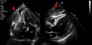
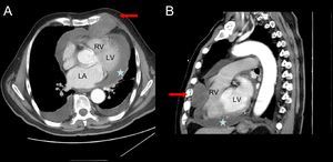
A 78-year-old man diagnosed with mitral valve prolapse and moderate mitral regurgitation presented to the emergency room with a 2-day history of edema and dyspnea. Echocardiography showed severe pericardial effusion with signs of increased intrapericardial pressure (Figure 1 and video of the Supplementary material; LA, left atrium; LV, left ventricle), as well as a large mass (Figure 1, arrows) adjacent to the free wall of the right ventricle (RV), in contact with the pericardium. Chest computed tomography showed a mass measuring 7×5×6 cm (Figure 2A and 2B, arrows) arising from the fifth costal arch and partially compressing the RV, as well as severe pericardial effusion (Figure 2, stars). The effusion was drained and showed no malignant cells, and the mass was removed; thereafter, the tumor was diagnosed as a low-grade chondrosarcoma. At 2 years following the procedure, the patient has had no recurrent disease.


The case presented is of interest because the mass was a slow-growing tumor that expanded toward the interior of the chest and manifested as an irritative pericardial effusion that established the diagnosis. This form of presentation is extremely uncommon. Chondrosarcomas of the chest wall are usually low-grade tumors with an excellent prognosis.