The patient was a 16-year-old boy who had been born with pulmonary atresia and ventricular septal defect, for which he had undergone complete repair. He had a Melody® pulmonary valve and stents in both pulmonary artery branches. He underwent placement of a dual-chamber cardioverter defibrillator for syncope and nonsustained ventricular tachycardia. The implantation was performed in the Department of Pediatric Cardiac Surgery. It was complicated by the tremendous complexity involved in placing the ventricular lead, due to inadequate capture and sensing (R wave) thresholds, making it necessary to evaluate several positions, until an apical position was chosen. Three months later, a very high ventricular capture threshold (6.5V × 1ms) and an R wave = 4mV were observed. Fluoroscopic examination ruled out lead displacement, and we were consulted with regard to its replacement.
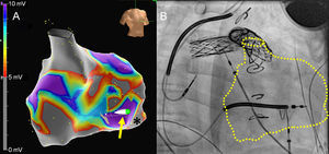
Given the difficulties of the initial implantation and to minimize the duration of the open surgery (due to the risk of infection), we decided to begin by constructing an endocardial voltage map of the right ventricle with the EnSite® Velocity® navigation system (St. Jude Medical) (Figure A, video of the supplementary material), using contact mapping with a sensor-equipped catheter to avoid recording floating points in the interior. After evaluating 203 points, we observed extensive regions with voltage < 5mV (grays), inadequate for implantation, and others ≥ 10mV that were suitable (violet). The previously implanted lead was in one of the gray regions (asterisk). The direct connection of the new ventricular lead to the navigation system itself made it possible to guide it for implantation in a healthy region. Figure A shows the final position of the lead (arrow), and Figure B corresponds to its fluoroscopic image. In this position, an adequate capture threshold (0.75V × 0.5ms) and an R wave = 9mV were achieved, and have been maintained after more than 1 month.
CONFLICTS OF INTEREST
J. Moreno has received fees for the development of presentations for Biosense Webster and St. Jude Medical.