Acute coronary syndromes and cardiogenic shock as a presentation of unruptured left sinus of Valsalva aneurysms have been described; however, this is the first reported case of a ruptured sinus of Valsalva aneurysm causing compression of the coronary arteries.
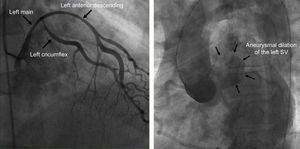
A 56-year-old man with a history of hypertension and tobacco use presented to the emergency department with chest pain after sexual intercourse. An electrocardiogram showed nonspecific ST-T wave changes in the inferolateral leads. The troponin I level was 3.14 ng/mL. Immediate coronary angiography demonstrated displacement of the left main, left anterior descending, and left circumflex arteries. The findings were suspected to be due to external compression by a hematoma from a type A aortic dissection affecting the left sinus of Valsalva (SV) (Figure 1).

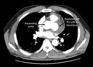
Computed tomography imaging revealed a large, partially thrombosed aneurysm of the left SV with a small hemopericardium (Figure 2), suggesting contained rupture of the left SV, which was causing compression and displacement of the coronary arteries.

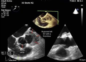
The patient was emergently taken to the operating room. A large fresh thrombus within the aneurysmal cavity was found and removed. The mouth of the aneurysm was approximately 3.0 x 1.5cm and leftward to the ostium of the left main artery (Figure 3). A vein graft bypass to the left circumflex, left internal mammary artery bypass to the left anterior descending, followed by a patch to the affected SV were performed. Forty days later, the patient was discharged home with a normal ejection fraction and neurological function.
