ISSN: 1885-5857 Impact factor 2023 7.2
Vol. 69. Num. 11.
Pages 1102 (November 2016)

Ecg contest
Response to ECG, October 2016

Respuesta al ECG de octubre de 2016

Francisco Buendía FuentesM. Asunción Hervás Botella
Rev Esp Cardiol. 2016;69:97810.1016/j.rec.2016.04.051
Francisco Buendía Fuentes, M. Asunción Hervás Botella

Options

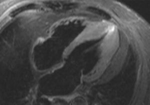
The clinical characteristics of pain, absence of ultrasensitive troponin I elevation, and absence of dynamic changes in the serial ECG make it unlikely that the patient has acute coronary syndrome (answers 1 and 2). This conclusion was supported by coronary angiography, which showed a disease-free coronary tree. The diagnosis was confirmed using cardiac magnetic resonance imaging (Figure), which revealed asymmetric apical ventricular hypertrophy (answer 3), as shown in the Figure. Edema, ischemia, and myocardial necrosis were ruled out, as well as pericardial disease (answer 4).

Figure
(0.06MB).

Some cases of apical hypertrophic cardiomyopathy can be overlooked in echocardiography,1 and ECG can show altered repolarization patterns,2 sometimes distinct from the typical T wave inversion in precordial leads.

References
[1]
R. Parisi, F. Mirabella, G.G. Secco, R. Fattori.
Multimodality imaging in apical hypertrophic cardiomyopathy.
World J Cardiol., (2014), 26 pp. 916-923
[2]
A.S. Flett, V. Maestrini, D. Milliken, M. Fontana, T.A. Treibel, R. Harb, et al.
Diagnosis of apical hypertrophic cardiomyopathy: T-wave inversion and relative but not absolute apical left ventricular hypertrophy.
Int J Cardiol., (2015), 183 pp. 143-148
Copyright © 2016. Sociedad Española de Cardiología
Are you a healthcare professional authorized to prescribe or dispense medications?