ISSN: 1885-5857 Impact factor 2023 7.2
Vol. 72. Num. 3.
Pages 248 (March 2019)

Ecg contest
Response to ECG, February 2019

Respuesta al ECG de febrero de 2019

Alejandro Cruz-UtrillaHernán Mejía-RenteríaAlejandro Travieso-González
Rev Esp Cardiol. 2019;72:16510.1016/j.rec.2018.04.032
Alejandro Cruz-Utrilla, Hernán Mejía-Rentería, Alejandro Travieso-González

Options

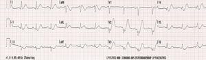
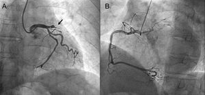
The electrocardiogram (ECG) (Figure 1), with ST depression and hyperacute T wave in precordial leads, allows the culprit vessel to be located (response 2, incorrect). As described by de Winter et al.1 in 2008, the ECG indicates acute proximal left anterior descending artery occlusion, as shown in the diagnostic coronary angiogram (Figure 2A, arrow), and so the correct response is answer 4. The ECG does not indicate a subocclusive lesion in the left main coronary artery (response 1, incorrect). In this case, the ECG would usually show diffuse ST depression and ST elevation in aVR.2 Likewise, this is not posterior ST-elevation acute coronary syndrome (response 3, incorrect). If it were, the ECG would possibly show ST elevation in the leads corresponding to the inferior face.

Figure 1
(0.47MB).
Figure 2
(0.2MB).

.

References
[1]
R.J. De Winter, N.J. Verouden, H.J. Wellens, A.A. Wilde.
Interventional Cardiology Group of the Academic Medical Center. A new ECG sign of proximal LAD occlusion.
N Engl J Med., (2008), 359 pp. 2071-2073
[2]
S. Kurisu, I. Inoue, T. Kawagoe, et al.
Electrocardiographic features in patients with acute myocardial infarction associated with left main coronary artery occlusion.
Heart., (2004), 90 pp. 1059-1060
Copyright © 2018. Sociedad Española de Cardiología
Are you a healthcare professional authorized to prescribe or dispense medications?