The ECG shows a tachycardia with a long RP interval (RP >PR) and negative P waves in the inferior leads and positive P waves in leads I and aVL. The differential diagnosis of long RP tachycardias includes orthodromic tachycardia with an accessory pathway, atrial tachycardia (AT), and atypical atrioventricular nodal reentry tachycardia (aAVNRT), but not typical AVNRT, characterized by short RP interval (RP< PR), and so option 2 is incorrect.1 Sinus tachycardia (option 4) can be ruled out because a positive P wave would be present in the inferior leads. AT is a possibility, but when this originates in the superior pulmonary vein, a positive P wave is usually present in leads II, III, and avF (option 1, incorrect).
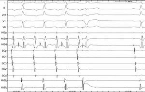
The correct response is option 3. An electrophysiological study confirmed the participation of a left posterolateral AP with decremental conduction in the tachycardia mechanism, given that the tachycardia terminated with ventricular extrasystole and the His bundle was refractory and without atrial activation (Figure 1).2 The tachycardia resolved with radiofrequency application at the AP site.
