Three-dimensional (3D) real-time echocardiography offers significant additional clinical information to traditional 2-dimensional echocardiography, improving visualization of cardiac structures and transcatheter device implantation.
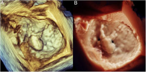
The recent development of high-definition photo-realistic rendering (EPIQ CVX, TrueVue, Philips Healthcare, Andover, USA) has been proposed as a new technological advancement aiming to increase the perception of 3D anatomical details, as in the case of degenerative mitral valve (MV) disease showing ruptured chords, height of the prolapse, and overriding of the posterior leaflet with respect to the anterior leaflet (Figure 1, and Video 1A and 1B of the supplementary data).

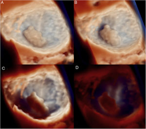
The technology allows users to change the lighting conditions to improve contrast. The light source can be moved around an annular plane to change the shadows of the MV (Video 2A and 2B of the supplementary data) or can be positioned at different heights with respect to a defined structure. In the case of MV analysis, the light source can be placed in the left atrium (Figure 2A), in the left atrium is just above the mitral annular plane (Figure 2B), in the left ventricle just below the mitral annular plane (Figure 2C), or deep in the left ventricle (Figure 2D).

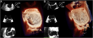
The photorealistic lighting effect has the potential advantage of improving our preoperative perception of cardiac anatomy (Figure 3A, and Video 3A of the supplementary data). It may also help in the assessment of morphological changes after cardiac procedures, such as transapical beating heart MV repair with neochord implantation showing a physiologic restoration of leaflet coaptation with simultaneous 2D-multiplane echocardiographic views (Figure 3B, and video 3B of the supplementary data).

In future, the clinical adoption of photo-realistic 3D echocardiography may improve our knowledge of this innovative technology, defining its real benefits and limitations.
Supplementary data associated with this article can be found in the online version, at https://doi.org/10.1016/j.rec.2014.04.021.