To the Editor,
.
Use of radial access has increased spectacularly. At the time of writing, it is found in 45% of all percutaneous coronary interventions in Spain.1 However, despite this, femoral access remains the most frequently used site in many catheterization laboratories. This is especially the case in North America and in patients in whom radial access cannot be canalized or when large caliber catheters are needed for complex procedures. We present the case of a patient with peripheral vascular disease and totally unsuitable for radial access who underwent coronary angiography and percutaneous intervention via a right axillo-femoral bypass.
A 61-year-old man with a history of smoking, high blood pressure, and hypercholesterolemia was admitted to a regional hospital with acute non-ST segment elevation coronary syndrome. In 2002, he had an aorto-bifemoral bypass for Leriche syndrome with juxta-renal obstruction of the aorta. Six years later he presented symptoms compatible with graft thrombosis. He was indicated for urgent arteriography and right branch thrombosis was confirmed. The patient received fibrinolysis, which was initially effective. However, when treatment ended he had another thrombotic episode leading to the decision for an urgent axillo-bifemoral bypass intervention that placed an 8mm polytetrafluoroethylene (PTFE) prosthesis from the right axillary artery to the deep femoral artery. At the time, coronary angiography was indicated at a regional hospital and the patient's clinical history showed the aorto-bifemoral bypass with no mention of the urgent axillo-femoral bypass. Echocardiography indicated normal ejection fraction. Radial access was attempted on both sides but the radial delivery catheter's hydrophilic guidewire could not advance. Given the existence of a very weak brachial pulse and good femoral artery pulse, coronary angiography was performed via the right femoral artery. The catheter was inserted with no complications and the PTFE prosthesis puncture dilated with a 6F introducer dilatation catheter (Cordis, Johnson & Johnson). The artery was canalized with the 6F delivery catheter and the guidewire and dilatation catheter extracted. When the JL4 6F catheter was advanced over the metal guidewire we discovered the patient had an axillo-bifemoral bypass, not just the aorto-bifemoral bypass described (Figure 1). We photographed the left coronary artery and a 285cm exchange guidewire was used to complete coronary angiography via the JR4 6F catheter. This showed no significant lesions in the left coronary artery and a long, severe lesion in the proximal and mid right descending coronary artery. Successful percutaneous placement of two drug-eluting stents was via a JR4 6F catheter guidewire similar to that used in femoral and radial access interventions. The procedure was completed with deferred manual extraction of the femoral introducer at 6h post-procedure. The patient was discharged next day with no complications.

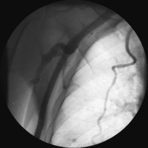
Figure 1. Contrast injection in right axillary artery. The brachial artery can be seen on the left. The axillo-bifemoral graft is in the center of the photograph and the right mammary artery is to the right.
Although percutaneous procedures on PTFE prostheses entailing puncture with the Seldinger technique have been described previously,2, 3 to our knowledge this is the first time this percutaneous coronary intervention has been performed via an axillo-bifemoral bypass. As radial and brachial access are possible on both sides, the need to resort to this access site can only arise in exceptional circumstances but may sometimes be of use. The procedure was conducted with standard percutaneous intervention materials with no difficulty. Hence, questions such as a theoretical limitation due to catheter length, difficulty in advancing through the graft, or lack of anatomy support, seem to be answered by the successful outcome of this case.
Corresponding author: inigo.lozano@gmail.com