Keywords
INTRODUCTION
The progression of heart failure is characterized by a series of compensation mechanisms in which hemodynamic, hormonal, and genetic factors modify the size, shape, and function of the ventricles.1 Despite the importance of the nucleus to cell function and of the nucleus-cytoplasm transport of macromolecules,2 their role in heart failure has not been studied.
The aims of the present study were to investigate the stereology of the nucleus (shape, size, and chromatin mass) in cardiomyocytes from patients with ischemic cardiomyopathy (ICM) and dilated cardiomyopathy (DCM), and to determine the expression of nucleoporin p62 (a molecule directly involved in nuclear transport3-5 through its association with the nuclear pore complex [NPC]) and its relationship with different nuclear variables.
METHODS
Collection of Samples
Experimental material was taken from explanted human hearts, 10 from patients with ICM (54 [4] years, all men) and 10 from patients with DCM (52 [6] years, again all men) (Table 1), all undergoing heart transplantation.

Clinical history, ECG, Doppler echocardiography, and coronary angiography data were available on all these patients; all had been classified functionally and all were receiving treatment for their conditions.
Transmural samples of tissue were taken at the apex and base of the left ventricle (maintained in NaCl 0.9% throughout the extraction procedure) and stored at 4oC for a maximum of 8 h from the time of coronary circulation loss. All samples were taken with the permission of the patients involved. The study was approved by the Ethics Committee of the hospital in agreement with the Declaration of Helsinki.
Optical Microscopy Analysis
The samples were fixed in a solution of glutaraldehyde (1.5%), formaldehyde (1%), and 0.05 mol cacodylate buffer (pH 7.4) for 60 min at 4oC, and then dehydrated and embedded in epoxy resin (Epon® 812). Semithin sections (50 µm) were stained with 0.1% toluidine blue and observed at a magnification of 60. Images were taken with a digital camera and stereological variables quantified using Scion Image Beta 4.02 software (Scion, MD, USA).
Electrophoresis and Western Blotting
Fifty milligrams of left ventricle were homogenized in a tube with sample buffer. These were then heated to 100oC, centrifuged for 5 min at 13 000 rpm (ambient temperature), and the supernatant distributed into aliquots for analysis. Their protein content was determined using the Lowry method. These samples were also subjected to 3%-8% gradient NuPAGE Tris-Acetate gel electrophoresis (Invitrogen). The proteins were then transferred to a nitrocellulose membrane (pore diameter 0.45 nm) and incubated for 2 h with rabbit polyclonal anti-goat IgG antibody (primary antibody, diluted 1/300; Santa Cruz Biotechnology, USA). Bands were visualized using an alkaline phosphatase-conjugated secondary anti-rabbit IgG antibody and a substrate system (Sigma Aldrich). Western blot quantification was performed using Scion Image Beta 4.02 software.
Statistical Analysis
Data are expressed as mean (standard deviation). The Kolmogorov-Smirnov test was used to examine the distribution of the variables (normal in all cases). Means were compared using the Student t test. Differences between percentages were analyzed using the Fisher exact test. Pearson correlation coefficients were calculated to determine the relationships between stereological variables and p62 expression. Multiple linear regression was then performed, using p62 expression as the dependent variable, and diagnosis, age, area of the nucleus, percentage heterochromatin and invaginated area (%) as independent variables. Significance was set at P<.05. All analyses were made using SPSS software for Windows v.11.0 (SPSS Inc.).
RESULTS
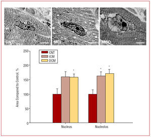
Images were obtained of 27 nuclei per heart (including controls; n=3). The cardiomyocytes from the patients with ICM and DCM showed larger nuclei and nucleoli - 60% and 66% larger respectively in ICM patients (P=.03 compared to the controls), and 59% (P=.02), and 75% (P=.03) larger respectively in the DCM patients (Figure 1). Small differences were also seen in the area of invaginated nuclear membrane, with a reduction of 5% in ICM patients and an increase of 11% in the DCM patients (compared to controls). Chromatin levels were similar in both disease states, with an increase of 4% and 6% compared to controls in the ICM and DCM patients respectively.

Figure 1. Optical microscopy sections of the left ventricle showing the area of the nucleus and nucleolus in ischemic cardiomyopathy (ICM), dilated cardiomyopathy (DCM) and in a control cell (CNT). Scale bar = 3 µm. The bar graph shows the relative areas of the nucleus and nucleolus in ICM and DCM cardiomyocytes compared to controls (represented as 100%). Values are mean (standard error). *P<.05.
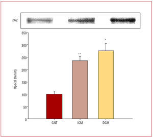
In the electrophoretic analysis, a significant increase in p62 was seen in the ICM setting compared to the controls (210 [48] compared to 100 [15] arbitrary units; P=.01). The same was seen in the DCM setting (245 [87] compared to 100[11]; P=.04) (Figure 2). No significant differences were seen between p62 expression in the patients of either disease group.

Figure 2. Western blot showing p62 expression in the control (CNT), ischemic cardiomyopathy (ICM) and dilated cardiomyopathy (DCM) settings. The data are expressed as the mean (standard error) of 5 independent experiments. *P=.04. **P=.01.
p62 expression was directly related to the area of the nucleus (r=0.615; P=.05) in the ICM patients. In addition, an inverse relationship was seen between p62 expression and the percentage of nuclear heterochromatin (r=0.707; P=.02 ) and the area of invaginated nuclear membrane (r=0.561; P=.09). In addition, a direct correlation was seen between the heterochromatin values and the area of the invaginated nuclear membrane (r=0.708; P=.005) (Figure 3). In the DCM setting, no significant correlations were seen. Finally, multiple linear regression showed p62 expression to be affected only by diagnosis (r2=0.46).

Figure 3.Correlations between nucleus area (A), percentage heterochromatin (B), and percentage invaginated nuclear membrane (C), and the expression of p62 in cardiomyocytes from patients with ischemic cardiomyopathy (ICM). Window D shows the correlation between percentage of invaginated nuclear membrane and the quantity of heterochromatin.
DISCUSSION
The results showed structural modifications to the nuclei of cardiomyocytes from patients suffering ICM and DCM, including significant increases in the areas of the nucleus and nucleolus. The size of the nucleolus is a direct reflection of its activity. In both patient groups the nucleoli were very enlarged since ventricular remodeling involves the increased expression of genes associated with cardiac adaptation mechanisms.6,7 However, no great differences were seen in the percentage of invaginated nuclear membrane, nor in the percentage heterochromatin in the cardiomyocytes of either type of patient.
p62 expression was significantly increased in both diseases. This may be a consequence of the activation of cardiomyocyte gene expression in heart failure and an accompanying increase in NPC-associated transport; this would lead to increases in the concentration of NPC components, including p62. A further consequence would be an increase in the size of the nucleus and a larger number of NPC in the nuclear membrane (note the correlation detected between the area of the nucleus and p62 expression).
When the nuclear stereological variables were compared with the expression of p62, significant correlations were obtained with the area of the nucleus, the percentages of heterochromatin, and the area of invaginated membrane, but only in the ICM patients. This could be due to the activation of different gene expression patterns in the two etiologies, which in turn activate different sequences of myocardial adaptation to biomechanical stress.7,8
A limitation of this work is its cross-sectional nature; no causal relationships can be established in this type of study nor can the response sequence be determined.
In addition, the small sample size limits any analysis by etiological subgroup, although clear correlations were obtained. Finally, since this is the first study of its type there are no data available for comparison.
In conclusion, patients with heart failure of ischemic or dilated etiology show modifications to the nuclear morphology of their cardiomyocytes and a large increase in the expression of p62 (which shows a relationship with changes in the size and shape of the nucleus in ICM). These changes may affect nuclear function in different ways in ICM and DCM patients.
ACKNOWLEDGEMENTS
The authors thank the Servicio de Coordinación de Trasplantes (Hospital Universitario La Fe, Valencia) for their help in obtaining the samples, and Inmaculada Montserrat (technician at the Laboratorio de la Unidad de Biología y Patología Celular del Centro de Investigación del Hospital Universitario La Fe) for her assistance.
ABBREVIATIONS
DCM: dilated cardiomyopathy
ICM: ischemic cardiomyopathy
NPC: nuclear pore complex
Correspondence: Dr. M. Portolés Sanz.
Unidad de Biología y Patología Celular. Centro de Investigación.
Hospital Universitario La Fe.
Avda. Campanar, 21. 46009 Valencia. España.
E-mail: portoles_man@gva.es
Received February 14, 2007.
Accepted for publication June 15, 2007.