These images belong to an 85-year-old man, with type 2 diabetes and no living ascendants or descendants, referred for evaluation after documentation of an exceptional cardiac morphology. He was in New York Heart Association functional class II and had frequent polymorphic ventricular extrasystole episodes.
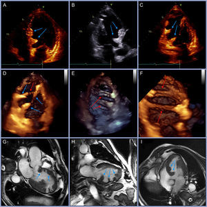
The 2-dimensional (Figure A-C) and 3-dimensional (Figure D-F) echocardiographic assessment revealed a dilated left ventricle with mild systolic dysfunction and multiple inwards digitiform images from the inferior and posterolateral walls and interventricular septum (blue arrows). Three-dimensional images particularly showed the circular ring arrangement of these projections and their apical linkage by several bands and false tendons, reflecting a particular myofibril architecture (red arrows). Cardiac magnetic resonance (Figure G-I) confirmed the presence of numerous digitiform images, without detection of the bands and false tendons identified on 2-dimensional echocardiography. There were no late enhancement findings or criteria for left ventricle noncompaction (characterized by the presence of a thin, compacted epicardial layer and an extensive noncompacted endocardial layer).

Myocardial perfusion scintigraphy showed ischemia, which did not correspond to epicardial disease on coronary angiography. The patient returned to New York Heart Association functional class I after beginning beta-blocker and renin-angiotensin-aldosterone system suppressing therapy, with better control of arrhythmias. Despite the uncertain prognosis of this cardiomyopathy, the patient had a relatively benign course with late detection and long survival.
This is a rare case of muscular dysplasia that has not yet been classified. In the few reports published to date, it is called sawtooth cardiomyopathy. After performing 3-dimensional echocardiography, we believe this designation to be unreliable. This imaging modality showed a complex intracavitary structure, with multiple digitiform images revealing several bands and false tendons, which translated into a deranged myofibril pattern.