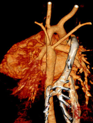
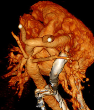
We report the case of a 30-day-old infant referred to our hospital with a diagnosis of anomalous right subclavian artery (ARSA) and suspected coarctation of the aorta based on echocardiographic findings. Prenatal ultrasound screening had detected the ARSA and the patient remained asymptomatic from birth with adequate weight gain. Physical examination revealed a systolic murmur, palpable pulses with no difference between upper and lower extremities, and absence of alterations on blood pressure registration. Electrocardiogram and chest radiography were normal. Transthoracic echocardiography showed mild hypertrophy of the left ventricle not affecting the ejection fraction, a bicuspid aortic valve, and dilatation of the ascending aorta. A continuous wave Doppler examination from the suprasternal notch showed a peak systolic pressure gradient in the descending aorta of 60mmHg with diastolic run-off indicating a severe obstruction at the classical site of a coarctation. Both subclavian arteries had a different origin than usual. Computed tomography (CT) confirmed the ARSA and coarctation of the aorta and demonstrated that subclavian arteries arose distal to the coarctation (Figure 1 and Figure 2), explaining the normal blood pressure in both arms. Because of the severity of the disease, the patient underwent cardiac surgery after diagnosis (reparation of the aortic arch and reimplantation of both subclavian arteries).
ISSN: 1885-5857
Impact factor 2023
7.2

