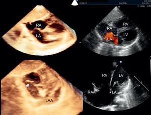
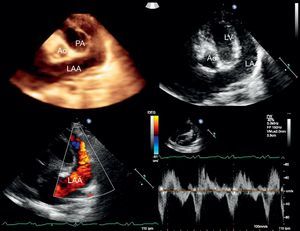
We present a case of left atrial appendage (LAA) aneurysm and atrial septal defect in a 1-year-old child without cardiovascular symptoms referred for cardiac murmurs and cardiomegaly and diagnosed by 2- and 3-dimensional transthoracic echocardiography. Subcostal and apical views showed a wide, fenestrated ostium secundum-type atrial septal defect with redundant septum (Fig. 1, arrow). The right-side cavities, including the right atrial appendage (RAA), were dilated. The LAA was prominent with a “small” left atrium (LA) (Fig. 1 and Video 1). Nonconventional slices showed the LAA aneurysm on either side of the upper pulmonary artery without causing an obstruction, surrounding the left ventricle (LV) at a lower level, and measuring 70mm; in the atrial ostium it measured 22mm, and in the middle section 20mm, without thrombus. Doppler images showed the auricular appendage interior flow emptying velocity was 60cm/s (Fig. 2, Videos 2 and 3). Biventricular function was conserved.


These aneurysms are rare, making diagnosis by echocardiography difficult because in the absence of knowledge about the predisposing heart condition they can be confused with other entities (eg, pericardial cyst, coronary artery aneurysm, left ventricular pseudoaneurysm, and congenital partial absence of pericardium). Three-dimensional echocardiography helps determine diagnosis as it shows the auricular appendage anatomy and its anatomic relationships, as does color Doppler, which shows filling from the LA. Transpulmonary contrast can also help define the atrial septal defect between the aneurysm and auricular appendage cavity.
The association with congenital heart disease is also infrequent. Patients can remain asymptomatic or be attended for arrhythmia or stroke. It has been suggested that the genesis of these aneurysms is in dysplasia of the pectineal muscle and the bands of connection in the auricular appendage.