We present the case of a 77 year-old woman with a past history of hypertension and permanent atrial fibrillation, who had an AAI pacemaker fitted via the right subclavian vein in 1980 for sinus node dysfunction. In 2009 she presented with atrial fibrillation with slow ventricular response and received a VVI pacemaker. Left subclavian access was used because the patient had right subclavian vein thrombosis, and the lead from the AAI was left in the right pectoral pocket. In December 2015, she was referred to our hospital; symptomatic failure to capture due to ventricular lead fracture at the sternoclavicular angle was recorded. Computed tomography angiography of the chest showed asymptomatic chronic obstruction of the superior vena cava; therefore, we decided to place a leadless pacemaker (Micra Transcatheter Pacing System) using right femoral access.
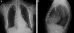
Chest X-ray (Figure A, posterior-anterior; Figure B, lateral) shows a remaining old lead in the right atrium running through the right subclavian vein and a remaining right ventricular lead placed via the left subclavian vein. The leadless pacemaker can be seen, along with the old ventricular lead, at the level of the right ventricular apex.

The new device was implanted with no immediate complications. Subsequent telemetry showed a pacing threshold of 0.25V at 0.4ms, impedance of 760 Ω, and an R wave of 5.2mV. The patient was discharged home after 24hours.
CONFLICTS OF INTERESTJ. Toquero is a member of Medtronic's European Advisory Board. Medtronic has sponsored his training in Micra implantation in Minneapolis, United States.