A 79-year-old man with a history of inferoposterior myocardial infarction and subsequent narrowing in the baseline electrocardiogram attended the clinic with progressive angina. Coronary angiography showed flow-limiting ostial stenosis of the first marginal artery and chronic occlusion of the second marginal artery. Single-photon emission computed tomography with methoxy isobutyl isonitrile (SPECT-MIBI) was requested with exercise testing to decide whether revascularization was indicated. This test was negative from the clinical point of view.
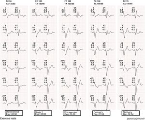
The electrocardiogram recorded during exercise testing is shown in figure 1.

What do you think was the most likely diagnosis?
- 1.
There is no cause for alarm, and the test should be considered negative.
- 2.
Spiked T waves are observed in the left precordial leads with exercise, indicating highly specific inducible ischemia.
- 3.
ST-segment depression was observed in the right precordial leads with exercise. This is a specific marker of inducible ischemia.
- 4.
An exercise-induced axis shift is observed. This is a highly specific marker of myocardial ischemia.
Submit your answer to http://www.revespcardiol.org/en/electroreto/73/02. The answer will be published in the next issue (March 2020). #RetoECG.