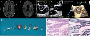
An 81-year-old woman presented to the emergency department with a transient episode of dysarthria and facial asymmetry. On cranioencephalic magnetic resonance imaging, multiple scattered areas of T2-weighted hypersignal and restricted diffusion were identified, being consistent with acute ischemic, possible embolic, lesions (Figure 1A and B, respectively). Both transthoracic and transesophageal echocardiography revealed several frond-like structures attached to the aortic side of the aortic cusps with no functional interference (Figure 1C, Video 1 of the supplementary data, 3D-multiplane). The differential diagnosis consisted of multiple valve tumor lesions, giant Lambl's excrescences, infective vegetations, despite negative blood cultures, and thrombi. Based on these findings, the patient underwent valve-sparing aortic valve surgery with an uneventful recovery. Multiple frond-like branched structures with distinct stalks were resected (Figure 1D). Histopathology provided the diagnosis of multifocal cardiac papillary fibroelastomas with multiple endothelial-lined fronds with a dense fibrovascular core and large amounts of elastic fibers (Figure 1E; HE, hematoxylin-eosin, Figure 1F; EvG: Elastic Van Gieson).

Unlike most cardiac tumors, papillary fibroelastomas cannot be appropriately characterized by advanced imaging tools such as cardiac magnetic resonance and computed tomography, due to their size, valvular cusp insertion, and significant mobility. Echocardiography not only identified several lesions but also allowed the planning of an aortic valve-sparing procedure. This was pursued as there were neither further structural abnormalities nor valve dysfunction. As large pedicles were identified, facilitating excision, and tumor recurrence is known to be rare, with few anecdotal, mostly metachronous tumors reported, the native valve was kept in place. Histopathology provided the definitive diagnosis, ensuring no need for further interventions.
Supplementary data associated with this article can be found in the online version, at https://doi.org/10.1016/j.rec.2018.11.014.