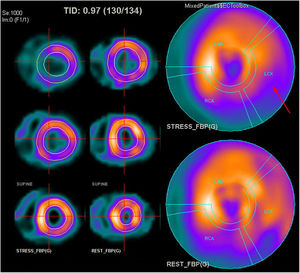
The electrocardiogram recorded during exercise testing shows a broadening of the final QRS complex over an underlying right bundle-branch block and left axis deviation (a left anterior hemiblock is present). This is indicative of inducible ischemia involving the anterior fascicle of the left bundle branch, and so response 4 is correct. Furthermore, after the performance of single-photon emission computed tomography with methoxy isobutyl isonitrile (SPECT-MIBI), reversible hypoperfusion is observed at the inferolateral wall, consistent with territory damaged by the known coronary artery disease (figure 1). Response 1 is incorrect because significant electrical abnormalities are present. Response 2 is incorrect because spiked T waves in the left precordial leads are a frequent finding in right bundle branch block, without necessarily indicating the presence of inducible ischemia. Response 3 is incorrect because, in presence of right bundle branch block, ST segment depression in the right precordial leads is frequent in the absence of inducible ischemia.
ISSN: 1885-5857
Impact factor 2023
7.2
