A 68-year-old man with an unremarkable history was admitted to hospital with a diagnosis of unstable angina. During his hospital stay, he experienced chest pain, with electrocardiographic changes on the inferior wall. Coronary angiography revealed a severe lesion in the middle third of right coronary artery, which was revascularized using a drug-eluting stent.
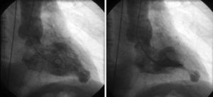
Ventriculography showed mildly depressed systolic function (ejection fraction, 54%), hypokinesis in the inferobasal region, and a saccular structure compatible with ventricular aneurysm in the apical region (Figure 1).

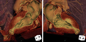
To characterize the findings, echocardiography and gated multidetector cardiac computed tomography (16 phases, 6.25% RR) were performed. A moderate-sized diverticulum was observed in the apical region of left ventricle (Figure 2; Video of the supplementary material).

Congenital ventricular diverticula are uncommon findings that are diagnosed incidentally, and are normally asymptomatic.
This structure can be viewed on echocardiography, but magnetic resonance is the ideal test to further define the diagnosis and course. In this case, given the experience and resources of the center, cardiac computed tomography was utilized.
The differential diagnosis should focus mainly on congenital or acquired ventricular aneurysms. The diverticulum has a narrow neck that communicates with the ventricular cavity, a wall composed of 3 differentiated layers, and systolic contraction synchronous with the ventricle. In contrast, aneurysms arise from a wide base. In histological terms, they lack a layer of myocardial muscle and have a single layer of fibroelastic tissue. During systole, they exhibit paradoxical expansion.
Cardiac computed tomography can be a useful and accurate tool in the diagnosis and characterization of ventricular diverticulum.