Cor triatriatum occurs during embryological development when the common pulmonary vein fails to incorporate in the left atrium (LA) and gives rise to a fibromuscular membrane that divides the LA into 2 chambers: a superior accessory chamber where the pulmonary veins enter, and an inferior one, the "true" LA, with its auricular appendage, which connects with the left ventricle through the mitral valve. Cor triatriatum is a rare congenital cardiac anomaly (0.4%).
A 46-year-old man consulted for palpitations and dyspnea, and atrial fibrillation was noted on the electrocardiogram. Cardiac catheterization was performed and the diagnosis of cor triatriatum was made on echocardiography findings.
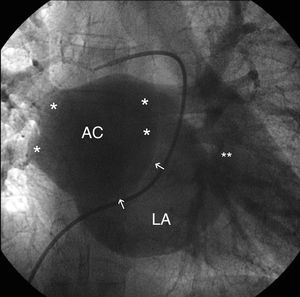
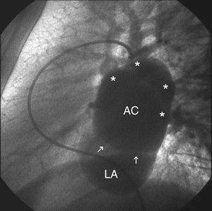
In the levophase of pulmonary angiography (Figures 1 and 2, anteroposterior and lateral views, respectively), an accessory atrial chamber (AC) connected to the 4 pulmonary veins (*) is observed, as well as considerable filling of the true LA through a fenestrated membrane (arrows). The LA contains the auricular appendage (**). No other connections were seen.

Figure 1.

Figure 2.
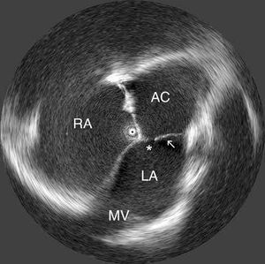
In the intracardiac echocardiography image (Figure 3), the transducer is seen in the right atrium (RA), the atrial septum is intact, and the accessory atrial chamber (AC) and true left atrium (LA) are clearly visualized, separated by an intra-atrial membrane (arrow) with a large filling orifice (*); the LA drains through the mitral valve (MV).

Figure 3.
This is a classic case of cor triatriatum, with an accessory chamber that receives the pulmonary veins and communicates with the LA.