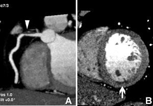
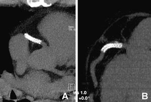
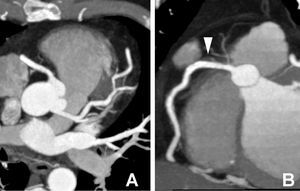
A 45-year-old male, smoker, and with no other cardiovascular risk factors was hospitalized for an inferior myocardial infarction. Fibrinolysis treatment was administered causing the clinical and electrocardiograph signs to disappear. A contrast-enhanced CT examination using a 16-multidetector CT was performed 3 days later. An atherosclerotic plaque with density measurements less than 50 HU (Hounsfield units) was observed in the proximal right coronary artery (RCA) (Figure 1A). Also, a small perfusion defect was observed in the inferior wall of the LV (Figure 1B). This plaque was classified as a complex lesion with 60% stenosis of the vessel lumen in a 60%. Several days later the patient presented with exertional angina and a SPECT was performed demonstrating a perfusion defect in the inferior myocardial wall with signs of viability. A coronary stent was implanted in the RCA. Two months later, when the patient presented with a typical chest pain a multidetector CT examination. The CT showed the coronary stent in a correct location (Figure 2A, 2B) with permeability within the lumen (Figure 3a, 3B). Thus, a conventional coronary angiogram was avoided.

Fig. 1.

Fig. 2.

Fig. 3.
Vulnerable plaques with a lipid-rich core can be demonstrated with this non-invasive imaging method, particularly when they have a density values less than 50 HU. In addition, in this case as in many others, multidetector CT can be used to evaluate the location and permeability of a coronary stent.