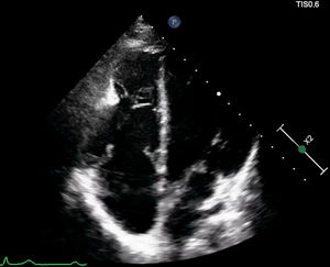
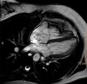
A 21-year-old woman with a previous history of patent ductus arteriosus (PDA) ligated in the neonatal age (thoracotomy) was referred to our department due to electrocardiographic anomalies. The patient remained asymptomatic and an electrocardiogram showed right precordial T-wave inversion, right-axis deviation, and normal progression of R-wave in precordial leads. Echocardiography showed mild right ventricle (RV) dilatation from different planes with bulging of the apical free wall, with no wall motion abnormalities (figure 1). Pulmonary pressure was normal and no residual PDA was detected. Cardiovascular magnetic resonance was performed to assess RV size and function, Qp/Qs, and to rule out arrhythmogenic cardiomyopathy with RV involvement. Heart displacement toward the left hemithorax with marked indentation of the RV mid-lateral wall was observed, suggesting partial herniation of the RV apical portion (figure 2). RV volumes and function were within normal values, and no regional wall motion contraction abnormalities were observed, thus excluding arrhythmogenic cardiomyopathy. Multiple sequences showed the absence of the parietal pericardium surrounding the mid-apical portion of the RV (figure 2). The discontinuity of the parietal pericardium, the left heart displacement, the bulging of the RV free wall, and the partial RV herniation was highly suspicious of partial absence of the RV pericardium. An echocardiogram in the neonatal age prior to PDA surgery revealed no pericardial anomalies. To avoid extra radiation in a young female, neither X-ray nor computed tomography was performed. Curiously, the RV showed a “heart-like” figure in the 4-chamber view (figure 2). To our knowledge, no previous case has reported both congenital anomalies occurring in the same patient.
ISSN: 1885-5857
Impact factor 2023
7.2

