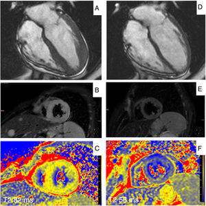
We present the case of a 49-year-old woman with recurring high-grade non-Hodgkin lymphoma and exercise-induced dyspnea, who was assessed for suspected apical hypertrophic cardiomyopathy following detection of symmetric negative T-waves in the anterior precordial leads in the ECG. Magnetic resonance imaging (steady-state free precession sequences) showed predominantly apical concentric left ventricular hypertrophy (figure 1A). Of note in the T2 weighted-Short Tau Inversion Recovery (T2w-STIR) sequences was a hyperintense signal (signal intensity ratio, 4), consistent with diffuse myocardial edema (figure 1B). Multiparametric tissue characterization enabled diagnosis of acute myocarditis with 2 major criteria according to the modified Lake-Louise criteria of 2018: increased T2-weighted relaxation time of 82ms in the mid level (regional normal value, 52±4ms) (figure 1C) and native T1-weighted relaxation time of 1.281ms (regional normal value, 995±36ms), with elevated extracellular volume of 32%. There was no late gadolinium enhancement.

A new magnetic resonance study was performed after immunotherapy with chimeric antigen receptor-T cell (CAR-T cell), revealing normalization of the wall thickness in the 17 left ventricular segments (figure 1D, end-diastolic cine sequence) and absence of signs of myocardial edema, with normalization of the T2w-STIR sequences (figure 1E) and parametric maps (figure 1F, mid-level T2 map).
This is the first published case of myocarditis associated with tumor relapse manifesting as transient left ventricular hypertrophy of apical predominance with ace of spades morphology, resembling cardiomyopathy. Such a presentation is associated with the possibility of diagnostic error. The case illustrates the importance of tissue characterization with multiparametric techniques for an appropriate diagnosis.