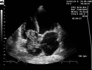
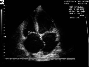
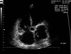
A 71-year-old woman with a history of right brachiocrural paresis occurring approximately 10 days prior to hospitalization, was admitted for repeated episodes of respiratory difficulty, with later development of jugular regurgitation, hepatomegaly, and edema of the lower limbs. In addition to these findings, the physical examination revealed a systolic murmur in the mesocardium. The baseline electrocardiogram showed atrial fibrillation. Doppler echocardiography, performed to assess ventricular function, revealed a large mass in the right atrium with a heterogeneous, multilobulated, mobile appearance, and a smaller mobile image in the left atrium, adhering to the interatrial septum (Figure 1, arrow). Anticoagulant treatment was started on the basis of suspected thrombus crossing a patent foramen ovale, together with an interatrial septal aneurysm. On serial echocardiographic controls (Figures 2 and 3), total disappearance of the right atrial mass was documented (Figure 3A), with persistence of the small mobile formation in the left atrium (Figure 3B). The presence of a patent foramen ovale was confirmed by color Doppler, which showed right-to-left flow and increased pulmonary pressure with respect to baseline values, as measured by the tricuspid reflux.

Figure 1. Baseline study.

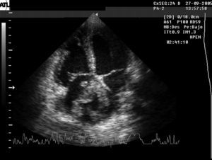
Figure 2. Study at 7 days.

Figure 3A. Study at 24 days, in which the interatrial septal aneurysm is evident.

Figure 3B. Study at 24 days. Persistence of the small image in the left atrium adhering to the interatrial septum.