Multimodality imaging plays an important role in preprocedural planning of transcatheter aortic valve implantation. A combination of multidetector computed tomography and transesophageal echocardiography (TEE) assessment is frequently used.
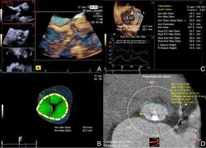
We used a recently commercially available 3-dimensional (3D) TEE probe (Z6Ms, Siemens Medical Solutions, Inc., Mountain View, California, USA) for aortic valve evaluation of a 95 year-old woman, referred to transcatheter aortic valve implantation for severe aortic stenosis (Figure A, Video 1 of the supplementary material). The eSie Valves software (Siemens Medical Solutions, Inc., Mountain View, California, USA) was used for the analysis (Figure B). Aortic annulus dimensions derived from TEE and multidetector computed tomography—using 3mensio software (3mensio Medical Imaging BV, Bilthoven, The Netherlands) —were compared.

Semiautomated TEE analysis showed a maximum and minimum annular diameter of 26mm and 19mm and a 72-mm perimeter (Figure C). By multidetector computed tomography evaluation, the maximum annular diameter was 26mm, the minimum was 18mm and the perimeter was 71mm (Figure D).
The 3D TEE transducer and eSie Valves software have recently been approved by the Food and Drug Administration. The probe allows 90°×90°real-time acquisition and full-volume color Doppler. The eSie Valves software provides rapid semiautomated valve measurements. This case suggests that the combination of a new 3D TEE probe and eSie Valves software may allow accurate measurements of the aortic valve annulus. To our knowledge, this is the first direct comparison between the 2 methods in preprocedural planning of transcatheter aortic valve implantation.