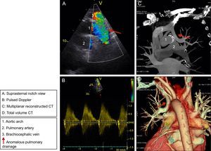
A 59-year-old woman was referred for paroxysmal atrial fibrillation. The physical examination revealed slightly elevated central venous pressure and cyanosis in the distal part of the phalanges, with no evidence of clubbing. Dilatation of the right cavities was observed in the chest X-ray. Two-dimensional transthoracic echocardiography confirmed this dilatation (video 1 of the supplementary material), with Qp:Qs of 1.3 and systolic pulmonary artery pressure of 85mmHg. The atrial septum was intact. In the suprasternal notch view (video 2 of the supplementary material and Figure A), systolic and diastolic flow acceleration was observed (pulsed Doppler, Figure B) in the region posterior to the aortic arch, behind the left subclavian artery. We suspected anomalous pulmonary venous drainage with the presence of a vertical vein draining into the brachiocephalic vein. The finding was confirmed by cardiac computed tomography (Figure C, multiplanar reconstructed computed tomography, and Figure D, total volume computed tomography).

Partial venous drainage of the pulmonary veins via the vertical vein is an uncommon condition that is observed in 0.5% of autopsies and occurs in 10% of anomalous drainage of left pulmonary veins. Given the limited clinical expression, this abnormality is underdiagnosed until adulthood.
This case shows that a meticulous echocardiography study, including a suprasternal notch view, may lead to diagnosis of this condition. Subsequently, cardiac computed tomography should be considered the method of choice to confirm the diagnosis.