Keywords
INTRODUCTION
The inability to reach peak heart rate during exercise testing is a factor that decreases the sensitivity and negative predictive value of myocardial perfusion single-photon emission computed tomography (SPECT) to detect ischemia.1-3 Thus, any method that helps to improve peak heart rate during exercise testing is of clinical interest.
Atropine is a muscarinic agent known to induce tachycardia and is commonly used in combination with dobutamine in echocardiographic and myocardial perfusion studies.4 However, its use as an aid in exercise testing has been little studied to date5-10 and its diagnostic value remains unknown compared to conventional stress protocols.
The study hypothesis was that scintigraphic criteria of ischemia during myocardial perfusion SPECT plus atropine obtained during submaximal exercise testing would be superior to those obtained with submaximal exercise testing alone. Thus, the aim of this work was to assess the diagnostic value of SPECT with intravenous atropine combined with submaximal exercise testing. This methodology could be used in clinical care during myocardial perfusion SPECT in patients who do not reach peak heart rate during exercise testing and do not have contraindications for atropine. It would be less difficult to administer (a perfusion pump would not be needed) and the duration of exercise testing would be reduced compared to other current pharmacological protocols (dipyridamole, adenosine, dobutamine).
METHODS
The ergometric and scintigraphic results were analyzed of a consecutive series of 172 patients who had been referred to myocardial perfusion SPECT for medical reasons and who had received atropine when they did not exceed 80% of their peak heart rate without symptoms of angina or ST-segment alterations.
Patients with the following contraindications for atropine were excluded: narrow angle glaucoma, obstructive uropathy, severe myasthenia, and obstructive gastrointestinal disease. In total, 23 of the patients who satisfied criteria for ischemia during SPECT underwent myocardial perfusion SPECT for a second time without atropine after 1 week with the aim of comparing the ergometric and scintigraphic results of the 2 tests. The study protocol was approved by the ethics committee of the hospital and all the patients gave their informed consent.
Exercise Testing Plus Atropine in Myocardial Perfusion SPECT
All patients underwent symptom-limited cycle ergometry testing (Biodex Medical System 8450A), with an initial 50 W load with 25-W increments per step every 3 min. A cephalic vein was cannulated before the procedure and 1 mg of atropine was administered intravenously to those who did not reach 80% of their peak heart rate without exhibiting angina or ST-segment depression ≥1 mm, while maintaining the maximum level of physical exercise they could tolerate for an extra 2 min. Between 30 s and 60 s before the end of exercise testing, 10 mCi of a radiotracer with technetium (methoxyisobutylisonitrile or tetrofosmin) were given by injection with the aim of acquiring tomographic images between 30 min and 60 min later. The device used was a dual-head 90o gamma camera (E.CAM, Siemens), equipped with low-energy, high-resolution collimators, set at 20 s/image with acquisition over a circular 180o orbit. After the images were acquired during submaximal exercise, 25 mCi of the same radiotracer were intravenously injected to obtain images during rest between 1 h and 2 h later.
Exercise Testing Without Atropine in Myocardial Perfusion SPECT
Within 1 week, 23 patients who satisfied scintigraphic criteria for ischemia during SPECT with atropine underwent exercise testing for a second time at the same level (power and metabolic equivalents [MET]) as before, but without atropine administration. Between 30 s and 60 s before the end of exercise testing, 10 mCi of the same radiotracer were administered to acquire exercise SPECT images. Resting images were not acquired, since the images obtained during the first study served as controls.
The following parameters were used to assess ischemia: clinical (angina), electrocardiographic (magnitude of horizontal or downsloping ST-segment depression) and scintigraphic (severity and extent of the perfusion defects). A 17-segment model was used in line with the American Heart Association guidelines,11,12 using a scale ranging from 0 points to 4 points according to the intensity of ischemia (0 = normal, 1 = slightly decreased uptake, 2 = moderately decreased uptake, 3 = severely decreased uptake, and 4 = absence of uptake). Without the observers being aware of the clinical and ergometric data or of the other test results, the scores of exercise SPECT with atropine, submaximal exercise SPECT without atropine, rest SPECT and the difference between the exercise and rest score (SDS) were quantified on the polar maps.13 Scintigraphic criteria for ischemia were satisfied when the SDS was ≥2.
Statistical Analysis
The SPSS version 15 statistical package was used. The clinical, electrocardiographic, and scintigraphic diagnostic variables of myocardial ischemia recorded in the first study (exercise with atropine) and second study (exercise without atropine) were compared. The results of the quantitative variables were expressed as mean (standard deviation) (range), whereas the qualitative variables were expressed as percentages or proportions. The normality of the data was determined using the Kolmogorov-Smirnov normality test. The Student t test was used to compare the quantitative variables of the 2 studies (exercise with atropine compared to exercise without atropine). The χ2 test was used to compare qualitative independent variables, and the McNemar test was used for the dependent variables. A P-value of <.05 was used as a cutoff for statistical significance.
RESULTS
The clinical characteristics of the 172 patients studied and of the 23 patients who underwent exercise testing with and without atropine are shown in Table 1. In total, 76 patients (44%) reached ≥80% of their peak heart rate and 113 (60%) a product heart ratexmaximum blood pressure ≥18 000. Heart rate and blood pressure significantly increased after atropine administration (Table 2). This increase could not have been due to the increase in the level of exercise, given that the load was not increased in any patient and even had to be slightly reduced in some patients so they could continue exercising for 2 min after atropine administration. There was a significant increase in heart rate in patients who were taking beta blockers (92 [13] vs 116 [19]; P<.0001). In total, 56 patients (32.5%) developed angina after atropine administration and 30 patients (17%) exhibited ST-segment depression ≥1 mm. No patient experienced unwanted adverse effects immediately after the administration of intravenous atropine and just 2 patients experienced nausea between 30 min and 60 min later.


The ergometric and scintigraphic results of the 23 patients who underwent SPECT with and without atropine are shown in Table 3. Twelve patients had previously undergone coronary angiography (6 had 3-vessel disease; 3 had 2-vessel disease; and 3 had 1-vessel disease). Of these 23 patients, 11 (48%) surpassed 80% of the peak heart rate. Six patients (26%) developed angina during the exercise test with atropine but not in the test without atropine. Furthermore, 5 patients (21.7%) exhibited ST-segment depression ≥1 mm in the exercise test with atropine but not in the test without atropine. In addition, 8 patients (35%) developed scintigraphic criteria of ischemia (SDS ≥2) during SPECT with atropine but not in SPECT without atropine (Figure 1 and Figure 2). The SDS was significantly greater in the SPECT studies with atropine (5.6 [4.5] vs 3.1 [2.8]; P=.0001).


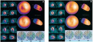
Figure 1. Examples of myocardial perfusion single-photon emission computed tomography (SPECT) with atropine (A) and without atropine (B) of a patient with stenosis of the anterior descending artery. The reversible septoapical defect observed during SPECT with atropine is not seen during SPECT without atropine. Quantification of the summed difference score (SDS) ranges between 4 and 1. SRS indicates summed rest score; SSS, summed stress score.

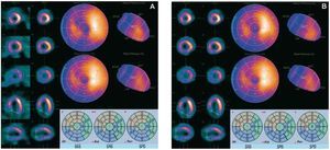
Figure 2. Examples of myocardial perfusion single-photon emission computed tomography (SPECT) with atropine (A) and without atropine (B) of a patient with inferior infarction and stenosis of the anterior descending artery. The partially reversible anteroapical defect is more evident during SPECT with atropine. Quantification of the summed difference score (SDS) ranges between 6 and 1. SRS indicates summed rest score, SSS, summed stress score.
DISCUSSION
When a patient is unable to perform any type of dynamic exercise, myocardial perfusion SPECT with the administration of dipyridamole or adenosine at rest14,15 or combined with isometric exercise16 can provide good sensitivity and specificity for the detection of coronary artery disease.17,18 Whenever a patient is able to perform dynamic exercise, the information provided by the clinical, hemodynamic and electrocardiographic parameters is of high prognostic value. For this reason, in practice, exercise testing is usually the first option proposed by the cardiologist as a provocation maneuver in bloodless diagnostic tests for ischemia.
Nevertheless, the sensitivity and negative predictive value of exercise testing and of myocardial perfusion SPECT are clearly related to maximum oxygen consumption and myocardial oxygen consumption.1 Thus, if 80% of the peak heart rate, product heart ratémaximum blood pressures of 18 000 and 5 MET cannot be achieved then the sensitivity and negative predictive values are prohibitively low.2 In these cases it has been demonstrated that dipyridamole3,19 or adenosine20 administration combined with physical exercise can significantly improve the diagnostic value of the test.
When conducting an ergometric test, the physician does not usually know if the patient will be able to reach peak myocardial oxygen consumption, especially peak heart rate, which is the determining parameter. Very often the inability of the patient to reach peak heart rate only becomes apparent during the exercise test itself when the patient is close to physical exhaustion. Dipyridamole administration at this point involves extending the exercise by an amount of time impossible for the patient to maintain, even if the load is reduced: 4 min of perfusion plus 2 min for administering the radionuclide and between 30 s and 60 s more before exercise is finally halted. The administration of atropine would make it possible to markedly shorten this extra time.
The intravenous administration of atropine has been combined with dobutamine with the aim of improving the diagnostic sensitivity of stress echocardiography4 and of myocardial perfusion SPECT21,22 when peak heart rate is not reached with dobutamine alone. Cousin-Sales et al6 administered 0.5 mg of intravenous atropine to 44 patients who underwent submaximal exercise testing, and obtained increases in maximum heart rate of 13.7 (7.4) beats/min; 74% reached a peak heart rate >80%. De Lorenzo et al7 obtained 85% peak heart rate in 44 of 47 patients (93.6%) who had received up to a maximum of 2 mg of atropine at the end of treadmill exercise testing. Our study hypothesis was to confirm whether intravenous administration of atropine to patients who do not reach a peak heart rate without clinical or electrocardiographic criteria for ischemia could improve the diagnostic yield of the test. Thus, this methodology was applied in a series of 172 patients in whom myocardial perfusion SPECT was indicated for diagnostic or prognostic reasons. No patient experienced unwanted adverse effects and 43.6% of the patients presented clinical (32.5%) or electrocardiographic (17%) signs of ischemia, and this would also increase the sensitivity of the conventional exercise test.
Furthermore, 23 of these patients with scintigraphic criteria for ischemia during SPECT with atropine underwent SPECT for a second time without atropine administration with the aim of comparing differences in clinical, electrocardiographic, and scintigraphic responses in the same patient.
We observed that when the patient is already unable to improve their level of exercise and the heart rate reached is submaximal, the administration of atropine at the end of exercise testing significantly increases heart rate and blood pressure. Thus, in almost one-third of the patients, there were scintigraphic signs of ischemia which would not have appeared without atropine administration. Furthermore, the severity of scintigraphic ischemia is significantly greater in studies in which atropine is administered.
Studies on the use of atropine combined with physical exercise during myocardial perfusion SPECT have demonstrated its safety, the absence of unwanted adverse effects and the capacity to reach 80%-85% peak heart rate in 74%-93% of patients, especially when beta blockers are being taken.6-8 Nevertheless, no study has analyzed the diagnostic value of myocardial perfusion SPECT in the same patients. In our series, we were able to verify that after atropine administration scintigraphic signs of ischemia appeared in up to 35% of cases which would not have appeared in the test without atropine. Furthermore, the severity of ischemia was significantly greater during SPECT with atropine.
The number of patients who underwent the 2 tests was limited as these tests involved patient irradiation, but we obtained statistically significant results and consider that the number of patients was sufficient to test the proposed hypothesis. Some authors8 have intravenously administered 0.5 mg of atropine every minute up to 2 mg if no clinical or electrocardiographic signs of ischemia appeared, although the average dose was 1 mg. For this reason, we administered the dose in a single bolus in our protocol, since this would not excessively prolong the test in some patients who were already on the threshold of physical exhaustion.
No studies have compared the diagnostic value of SPECT plus atropine during submaximal exercise testing to SPECT plus dipyridamole during submaximal exercise testing in the same patient; thus, this methodology cannot be recommended in all patients who do not reach peak heart rate. Nevertheless, atropine administration could be considered in patients who are very close to physical exhaustion during exercise testing, if the cardiologist considers that they cannot continue with the exercise for the time needed to proceed to dipyridamole perfusion. Future studies could compare the diagnostic value of SPECT with atropine during submaximal exercise testing to this value when using other vasodilators in the same patient.
Thus, SPECT with intravenous atropine combined with physical exercise could be used in clinical care in patients who do not reach peak heart rate during exercise testing and do not have contraindications for the drug, since this involves less complexity (no perfusion pump is needed, in contrast to adenosine) and less exercise time compared to the exercise plus dipyridamole protocol.3
CONCLUSIONS
Our results indicate that one-third of patients who met scintigraphic criteria for ischemia at the end of submaximal exercise testing and after atropine administration would not have met those criteria without administration of the drug.
ABBREVIATIONS
IV: intravenous
MET: metabolic equivalent
SDS: summed difference score
SPECT: single-photon emission computed tomography
SRS: summed rest score
SSS: summed stress score
This study was partially financed by a grant from the REdes temátiCAs de investigación cooperatiVA, Carlos III Institute (Red C03/01, RECAVA).
Correspondence: Dr. J. Candell Riera.
Servei de Cardiologia. Hospital Universitari Vall d'Hebron. Passeig Vall d'Hebron, 119-129. 08035 Barcelona. Spain.
E-mail: jcandell@vhebron.net
Received January 21, 2009.
Accepted for publication March 24, 2010.