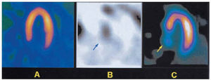

Fig. 1
A 62-year-old patient had symptomatic hypertrophic obstructive cardiomyopathy diagnosed six years ago. Despite medical treatment, dyspnea progressed during follow-up and a two-chamber pacemaker had been implanted one year previously. The reason for the current hospitalization was worsening symptoms (functional class III). The dynamic left ventricular outflow tract gradient was 120 mm Hg by Doppler. Single photon emission computerized tomography (SPECT) myocardial perfusion images showed no perfusion defects (Figure 1A). The patient's symptoms were refractory to treatment, and percutaneous alcohol septal ablation was recommended. Alcohol injection into a dominant third septal artery branch resulted in septal myocardial infarction. There were no complications and the intraventricular dynamic gradient decreased to 20 mm Hg. Cardiac scintigraphy with 99mTc-pyrophosphate performed on the third day after ablation showed an area of intense septal uptake (Figure 1B, arrow). In resting SPECT images with 99mTc-tetrofosmin done on the fifth day post-ablation, reduced uptake was visualized in the basal septal region (Figure 1C, arrow), corresponding to the area of necrosis. The patient's subsequent clinical course was satisfactory.
Although the clinical, echocardiographic, and hemodynamic results of percutaneous septal ablation in hypertrophic obstructive cardiomyopathy have been the subject of several publications, radionuclide scintigraphic images of this therapeutic procedure have not previously been published.