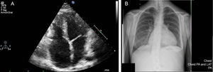
The echocardiogram (Figure A) shows a nondilated mildly hypertrophic left ventricle with preserved contractility, significant biatrial dilatation, a type I diastolic dysfunction mitral flow pattern, and significant dilatation of the suprahepatic veins, all of which is compatible with a diagnosis of restrictive cardiomyopathy. Right heart catheterization confirmed the restrictive physiology.

Copyright ©
2015. Sociedad Española de Cardiología