
A 65-year old woman with a prior history of surgical mitral comissurotomy 30 years ago presented with a 6-month history of worsening fatigue and exertional dyspnea. On physical examination, she had a mitral stenosis murmur, a loud P2, with an irregular pulse and mild peripheral edema. The electrocardiogram showed atrial fibrillation; the echocardiogram was notable for a fibro-calcified mitral valve, with severe restriction of leaflet mobility and an area of 1.1 cm2. Biventricular function was normal.
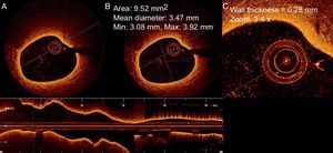
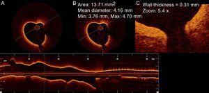
As the valve anatomy was considered suitable for percutaneous intervention, the patient was scheduled a balloon valvuloplasty. The pre-intervention right heart catheterization showed a pulmonary artery pressure of 45/17/29mmHg. After successful mitral dilatation, optical coherence tomography (LightLab Imaging Inc., Westford, Massachusetts, United States) was performed on a distal segmental branch of the right pulmonary artery (Fig. 1).

Optical coherence tomography images showed diffuse thickening of the distal pulmonary arterial wall (Figs. 2 and 3). We registered a pulmonary artery wall thickness between 0.28mm and 0.31mm, higher when compared to reports from subjects without pulmonary hypertension (0.16 [0.03] mm for vessels with 2.14 [0.33] mm of diameter). No complications arose during or after the procedure.


Optical coherence tomography is a safe and potentially useful tool for characterize, with high resolution, the pulmonary vessels and may contribute to investigate the mechanisms of vascular remodeling in pulmonary hypertension.
Acknowledgments: We are indebted to Mark Green, Paula Lopes, Cristina Silva, and Joana Ferreira.
CONFLICTS OF INTERESTDr Jorge received a research grant from St Jude Medical, Portugal.