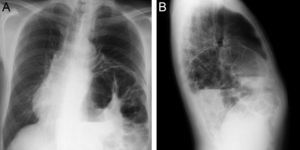
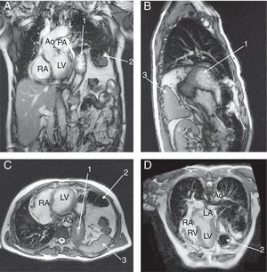
A 63-year-old male patient underwent a routine medical check-up. Physical examination revealed the presence of muffled heart sounds, absence of palpable apical impulse, and decreased breath sounds on the lower left side of the chest on auscultation. Moreover, gurgling sounds suggestive of bowel sounds were audible in the left side of the chest on auscultation. Abdominal examination was unremarkable. Chest X-ray showed a shift of mediastinal structures to the right because of the presence of incidental herniated bowel loops in the left hemithorax with several air-fluid levels (Fig. 1). A magnetic resonance imaging study of the thorax was performed (Fig. 2A coronal, Fig. 2B sagital, Fig. 2C axial, and Fig. 2D cardiac 4-chamber plane; Ao, aorta; PA, pulmonary artery; RA, right atrium; LA, left atrium; RV, right ventricle; LV, left ventricle). Magnetic resonance imaging demonstrated normal visceral situs, a compressed left lung, cardiac displacement to the right, and levo-orientation due to the existence of a large, left posterior diaphragmatic hernia. The stomach, spleen, and left colon were displaced into the left thoracic cavity through the posterior diaphragmatic defect (indicated as 1, 3, and 2 respectively, in Fig. 2). Left diaphragmatic hernia (Bochdaleck hernia) is a cause of cardiac dextroposition or pseudodextrocardia, which is defined as a displacement of the heart to the right secondary to extracardiac causes. Despite being well informed about the potential complications of noncorrected diaphragmatic hernia, the patient refused surgery.
ISSN: 1885-5857
Impact factor 2023
7.2

