
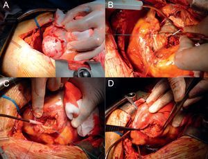
A 74-year-old woman with endocarditis of a mechanical mitral valve was referred for coronary angiography prior to valve replacement surgery. Investigations revealed a coronary tree without atheromatosis, systolic tapering of the distal circumflex artery after the exit of the atrioventricular groove and transitory occlusion of the vessel, indicating extrinsic compression at this level (Figs. 1A and B). Ventricular angiography showed retention of contrast in a cavity posterior to the left ventricle, with clearly defined borders and a narrow entrance (Figs. 1C and D). Surgery was performed, during which a giant ventricular pseudoaneurysm was found in the posterior wall of the left ventricle (Fig. 2A) with entry site at the level of the posterior mitral annulus (Fig. 2B, arrow), which was thoroughly dried, with defect closure using a Dacron patch (Figs. 2C and D).


Pseudoaneurysms of the left ventricle posterior wall are extremely rare and generally develop after myocardial infarction in this territory. Other causes include postsurgical rupture of the posterior annulus, congenital weakness of the posterior wall, thoracic trauma or infective endocarditis. In our patient, there were 2 possible coinciding etiologies: postsurgical rupture of the annulus and the presence of infective endocarditis. The surgical findings and history of prior surgery make rupture of the posterior annulus the more likely cause. An image of “milking” or atypical coronary systolic compression should lead to suspicion of this entity.