
A 65-year-old woman attended the cardiology service with dyspnea and presyncope. The patient's medical history included Hodgkin lymphoma treated in her youth with chemotherapy and radiotherapy and breast carcinoma treated by surgical resection, radiotherapy, and hormone therapy. She was under treatment at the cardiology service for moderate mitral and tricuspid regurgitation with preserved biventricular function, attributed to her history of radiotherapy. An echocardiography examination excluded progression of the valve disease.
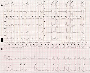
An electrocardiogram (figure 1) revealed sinus tachycardia versus a perisinus node atrial tachycardia of 140 lpm (asterisks) with variable atrioventricular conduction. Intermittent conduction via the left anterolateral accessory route (figure 1, solid arrows) alternated with episodes of complete atrioventricular block with escape beats with a right bundle block pattern (figure 1, dashed arrows).

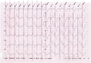
A dual chamber pacemaker was implanted. The postprocedure electrocardiogram (figure 2) showed a wide-QRS sinus rhythm with 2 alternating patterns. One pattern corresponded to pacemaker-stimulated ventricular beats (figure 2, dashed arrows), the other to conduction via the accessory route with a pacemaker spike in the initial portion of the QRS complex of the pseudofusion beat (figure 2, solid arrows). An alternative explanation would be the presence of ventricular extrasystole of the anterolateral mitral annulus; however, this is less likely in the context of a constant PR interval and variable couplings.

The electrocardiogram is an essential tool and should be analyzed in detail. Not all pacemaker spikes preceded by the start of the QRS should be considered a sensing failure. On this occasion, the spike coincided with pseudofusion beats. The findings were confirmed by electrophysiological analysis. Informed consent was obtained for publication.
FUNDINGThe present study did not receive funding.
AUTHIORS’ CONTRIBUTIONSAll authors contributed equally to the article.
CONFLICTS OF INTERESTThere are no conflicts of interest to declare.