To the Editor:
The radial artery is widely recognized as an excellent access route for diagnostic and interventional coronary procedures; many interventional cardiology units consider it the access of choice in patients with no contraindications. The route is safe and has a low incidence of complications.1,2 In 2005 more than 30 000 procedures were performed in Spain using the radial approach, 56% more than in the previous year.3 Anatomical variations that hinder the procedure are not rare, but can be solved using long hydrophilic guidewires or introducers.4 Severe peripheral arterial disease, although encountered less often, may require switching to another route. An alternative could be interventional treatment of the lesion, which would allow the procedure to be continued.
We describe an 83-year-old man with hypertension, diabetes, and Fontaine stage IIb intermittent claudication. Relevant history included remote inferior myocardial infarction documented by electrocardiography. The patient was referred for non-ST-segment-elevation acute coronary syndrome with elevated markers of myocardial injury. An Allen test performed before the procedure was normal.
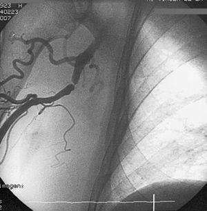
The procedure was initiated using the right radial approach, with an Avanti Plus 6F hydrophilic introducer (Cordis Corp., USA); diagnostic angiography was started with a JL 3.5 catheter and 0.38-inch guidewire. Because it was difficult to advance the guidewire near the axillary region, a control injection was performed. The brachial artery showed a severe focal stenosis after the origin of the humeral circumflex artery, with large-diameter collateral circulation to the deep brachial artery that ensured blood supply to the distal limb and explained the absence of symptoms (Figure 1). After characterizing the lesion in several planes and injecting 200 µg of nitroglycerin, a decision was made to intervene.

Figure 1. Diagnostic angiography.
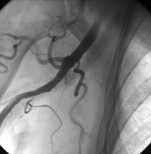
The introducer was exchanged for a 7F device and the lesion was crossed with a BHW 0.014-inch intracoronary guidewire (Guidant Corp., USA), reaching the aortic valve plane. Predilation was performed with a 320-mm Powersail noncompliant coronary dilatation catheter (Guidant Corp., USA) at a pressure of 12 atm, checking for expansion. Lastly, a 516-mm Titan2 stent (Hexacath, France) was implanted, with complete resolution of the stenosis (Figure 2).

Figure. 2. Final outcome.
Once the brachial artery stenosis had been dilated, diagnostic angiography was continued using the same route without complications. Chronic occlusion was found in the distal right coronary artery and angiographically nonsignificant stenosis in the left anterior descending artery; hence, the patient was referred for a more complete ischemia and viability study. The amount of contrast used was 300 mL and imaging time was 24 min. The patient was transferred to the respective reference hospital at 24 h, with no complications and with dual antiplatelet therapy for 1 month.
Percutaneous surgery in the subclavian and vertebral arteries or the brachiocephalic trunk has been described in the case of vertebrobasilar insufficiency.5 However, there are no relevant publications on percutaneous treatment of the brachial artery during coronary angiography. This situation could change in upcoming years as the radial approach is gradually implemented for access in coronary angiography and because of the increasing comorbidity of treated patients.
In these cases, there are 2 possible approaches: to change the access route or to treat the lesion. The decision must be made on a case-by-case basis, assessing potential complications. In the patient presented, the location of the occlusion and the collateral circulation that ensured distal flow made the surgery safe, and the approach was then suitable for later potential use in any coronary revascularization procedure.