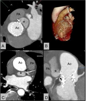
A 77-year-old man presented with progressive chest pain and ST-segment depression. He had undergone transcatheter aortic valve implantation (TAVI) with a 34-mm CoreValve Evolut R (Medtronic, United States) implanted in another center 6 months previously. A transesophageal echocardiogram revealed a large cavity connected to the ascending aorta, with active flow inside. An urgent computed tomography showed a large pseudoaneurysm of the ascending aorta between the pulmonary artery (PA) and the aorta (Ao) (figure 1A-D, asterisk) with a narrow neck (figure 1A, white arrow), suggesting compression of the left main (LM, black arrow) (figure 1C,D). The case was discussed in a medical-surgical session and it was decided to perform immediate percutaneous repair.

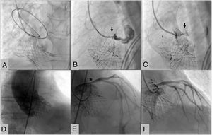
From right radial access, a hydrophilic guidewire with support with a JR 4 guiding catheter was advanced into the pseudoaneurysm until various loops formed (figure 2A). An 8-mm Amplatzer Vascular Plug II was subsequently implanted, with progressive closure of the cavity in the next minutes (figure 2B-D, arrow, video 1 and video 2 of the supplementary data). Left coronary angiography showed a fixed stenosis of the proximal LM (figure 2E, asterisk), which was treated by implanting a stent with a good result (figure 2E,F).

Pseudoaneurysm of the ascending aorta is a potentially lethal complication normally arising after interventions involving surgical manipulation of the aorta. Although the certain origin in this case is unknown, it probably corresponds to a contained aortic rupture unnoticed during valve implantation, which progressively expanded. Percutaneous closure of aortic pseudoaneurysms is a safe and effective intervention. An exhaustive study of the anatomy is recommended in order to design a tailored strategy for this kind of procedure.
Supplementary data associated with this article can be found in the online version available at https://doi.org/10.1016/j.rec.2020.10.005