
Ventricular noncompaction is a recognized variety of myocardial disease involving systolic and diastolic ventricular dysfunction.
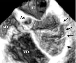
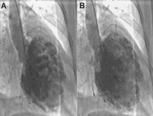
We present a 3-D echocardiographic reconstruction in a 52-year-old woman with a 2-year history of palpitations and heart failure that improved with initiation of pharmacologic therapy. The electrocardiogram showed sinus rhythm at 70 bpm with a first-degree atrioventricular block and indeterminate axis, with evidence of left atrial enlargement and dilation of the left ventricle with diffuse repolarization disorders (Figure 1). The electrophysiological study showed severe sinus node and atrioventricular dysfunction; non-sustained ventricular tachycardia was induced by stimulating the right ventricular outflow tract. The transthoracic echocardiogram showed noncompacted myocardium and ventricular dysfunction, and 3-D transesophageal echocardiography in the four-chamber view demonstrated myocardial noncompaction (Figure 2). The arrows in the image indicate the region of noncompacted myocardium, which is seen as multiple bands extending from one side to the other of the left ventricular chamber (RA indicates right atrium; LA, left atrium; Ao, aorta). Recording of pressures with cardiac catheterization confirmed the diastolic ventricular dysfunction. Left ventriculography in the right anterior oblique view in systole (Figure 3 A) and diastole (Figure 3B) showed dilation and severe generalized hypokinesia of the left ventricle with multiple trabeculations.

Fig. 1.

Figure 2.

Figure 3.