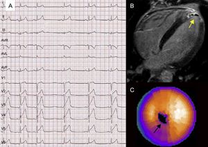
A 34-year-old male smoker with a family history of sudden cardiac death, consulted for pleuritic chest pain. ST-segment elevation (Figure 1A) with elevated troponin I concentration and normal echocardiography findings led to a diagnosis of myocarditis.

Magnetic resonance imaging showed late enhancement in the distal anteroseptal segments and apex, indicating infarction, with internal hypointensities consistent with small areas of bleeding within the infarction (Figure 1B, arrow). Technetium-99m sestamibi single-photon emission tomography showed an absence of viability in the apex (Figure 1C, arrow).
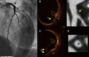
Coronary angiography detected a nonsignificant plaque in the distal trunk (Figure 2A, arrow), characterized by optical coherence tomography as an eroded plaque (Figures 2B and 2C, asterisk) with a feature indicating an adhered thrombus (Figure 2B and 2C, arrows). A 64-row multidetector computed tomography examination also detected the plaque, which was nonobstructive and hypoattenuated (Figures 2D and 2E, arrow), with some positive remodeling and a hyperattenuated ring surrounding the hypodense plaque (napkin ring sign).

The patient was discharged with antiplatelet therapy (aspirin + ticagrelor) and a diagnosis of apical infarction due to embolism of an eroded, nonobstructive plaque in the distal trunk.
The differential diagnosis between myocarditis and infarction is often a challenge. Magnetic resonance imaging provides the greatest diagnostic yield in these patients. Plaque erosion is the underlying cause of 30% of ST-elevation myocardial infarctions and may go unnoticed on coronary angiography. Optical coherence tomography would be the ideal technique to establish the etiological diagnosis in patients with infarction and no obstructive lesions. Multidetector computed tomography can identify characteristics of the plaque indicating vulnerability to rupture (eg, positive remodeling, hypoattenuation, napkin ring sign) and could be useful for follow-up purposes.