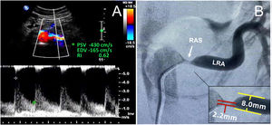
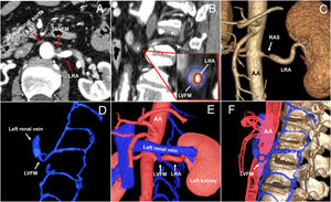

A 64-year-old male patient was admitted to hospital due to high blood pressure with recurrent dizziness and headache for the previous 2 months. Laboratory test results showed normal catecholamine hormones, adrenocorticotropic hormone and cortisol determination and a high level of renin assay (upright renin, 500.00 uIU/mL; recumbent position renin, 164.89 uIU/mL). Ultrasound examination (figure 1A) of the renal artery revealed that the left renal artery (LRA) was narrowed at the beginning with peak velocity of 430.0cm/s. The diameter of the renal artery stenosis site was 2.2mm measured on digital subtraction angiography (figure 1B). Renal artery computed tomography angiography (CTA) examination showed a venous fenestration malformation wrapped around and constricting the LRA (figure 2A,B; videos 1-3 of the supplementary data), while the kidneys and adrenal glands were normal, and no plaque was found in bilateral renal arteries. Volume rendering (VR) and multistage vascular fusion technology (figure 2C-F; video 4 of the supplementary data) visually displayed that the lumbar venous fenestration malformation (LVFM) was enveloped and constricted the beginning of LRA, close to the abdominal aorta. After clarification of the cause of hypertension, the patient refused surgery. Unfortunately, he died due to hematencephalon 1 week after discharge. Written informed consent was obtained from the patient's wife for publication of this case report and accompanying images.


Renal artery stenosis (RAS) is a common cause of secondary hypertension. This is the first report of RAS due to LVFM. CTA and VR can visually display this rare cause of RAS.
FUNDINGInner Mongolia Autonomous Region Higher Education Research Fund Project [grant number NJZY21082].
AUTHORS’ CONTRIBUTIONSR. Zhang compiled the data, interpreted the computed tomography image of the renal artery, and wrote the article. Y. Zhao provided a critical review of the article. Q. Ji interpreted the computed tomography image of the renal artery and provided a critical review of the article. All authors approved the final version.
CONFLICTS OF INTERESTNone declared.
Supplementary data associated with this article can be found in the online version, at https://doi.org/10.1016/j.rec.2022.07.006