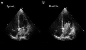
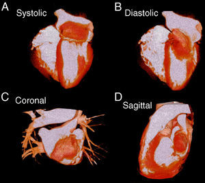
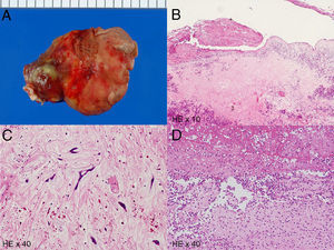
A 62-year-old man with a history of diabetes was admitted to the hospital following complaints of fever and fatigue. The patient was asymptomatic until 2 weeks prior to admission, when the above symptoms developed. Specimens of blood culture yielded Streptococcus agalactiae and a left atrial tumor was detected by cardiac ultrasound (Fig. 1). Even with the use of intravenous antibiotics therapy for 2 weeks, the patient still had a fever. He was transferred to our institution to undergo further testing and treatments. Transesophageal echocardiography showed no valvular vegetation, but revealed a mass adhering to the left atrial septum. A 320-slice dynamic volume multislice computed tomography scan (Toshiba Aquillion One, Tokyo, Japan) was performed to evaluate coronary artery disease and cardiac tumor before the surgery. It confirmed no significant coronary artery narrowing and the presence of the left atrial mass (61×21mm) with vegetations (Fig. 2). Surgical removal of the myxoma was thus performed the day after admission. The tumor has a stalk and is covered with thrombi (Fig. 3A). A pathology examination confirmed the mass to be an infected myxoma (Fig. 3B). The tumor consisted of an acid mucopolysaccharide matrix and polygonal cells, and the tumor surface showed neutrophils, fibrin, and thrombi (Figs. 3C and D). The patient subsequently underwent antibiotic therapy for 4 weeks and was then discharged. At the end of 1 year, he remained asymptomatic and displayed no evidence of recurrence on echocardiography.


.