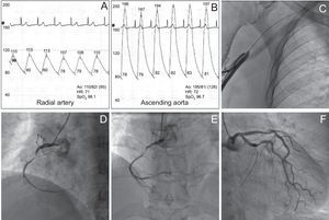
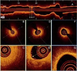
A 49-year-old woman with Takayasu arteritis was referred for coronary angiography after having a non-ST elevation acute inferior myocardial infarction. The right transradial approach revealed elevated central blood pressure with normal peripheral blood pressures, near occlusion of the axillary artery (Figures 1A-C), and severe stenosis in the middle portion of the right coronary artery (Figures 1D-F and Video 1 of the supplementary material). Optical coherence tomography (OCT) was performed in the culprit lesion. At the critical level of the stenosis, there was severe fibrointimal thickening with apparent endothelial integrity and no evidence of thrombus (Figures 2A-C and Video 2 of the supplementary material). Distal to these findings, a region of heterogeneous, hypointense signal with well-defined margins indicated a focal calcium deposit (Figures 2D-E). Proximal and distal to this observation, there were infiltrated hyperintense areas of asymmetric fibrointimal thickening with an acoustic shadow indicating macrophages (Figures 2E-G). Finally, the patient underwent stent implantation (Video 3 of the supplementary material).


Multifactorial cardiac involvement is the major cause of death in Takayasu arteritis. Our understanding of the underlying pathophysiology of the coronary artery involvement is based mainly on autopsies. The spatial resolution and in vivo nature of OCT provide a unique opportunity to study the virtual histology of Takayasu arteritis and broaden our knowledge of this entity. In contrast to an earlier case in which only fibrous scar tissue was observed in a patient with ventricular dysfunction, in our patient, OCT demonstrated a critical stenosis associated with a calcium deposit as the underlying pathophysiology of the acute coronary syndrome, in addition to patchy involvement due to a chronic active inflammatory process in the arterial wall.