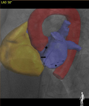
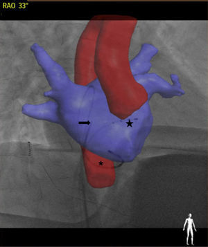
In the setting of electrophysiology, transeptal puncture is a key phase during numerous procedures. This technique is not risk-free, and certain measures should be applied to minimize them. In the present case, we attempt to describe how integration of the scanner with the intraprocedure radiologic image is feasible with the new rotational angiography systems (Philips Allura FD20 EP Navigator 3-dimensional angiography), in order to increase the safety of the technique. Once computed tomography (CT) is incorporated into the system, automatic 3-dimensional segmentation of the different structures can be performed. Subsequently, the 3-dimensional volume is superimposed on the fluoroscopy image (hence, 3 views separated by more than 30° should be used) and the overlay and image are matched using a fixed anatomic landmark as reference (eg, the carina). The following images illustrate how it is possible to define the aorta and atria and visualize the true position of the catheters. It may also be useful to localize the fossa ovalis. Figure 1 shows a left anterior oblique view, and Figure 2 a right oblique view, which perfectly correlate with the bundle of His catheter (which marks the level of the aortic valve) and coronary sinus (atrioventricular groove) (star and asterisk, respectively). The Brockenbrough needle emerges in the middle through the Mullins sheath (arrowhead). The technique is so highly accurate that arterial or venous punctures to delineate the aorta or bundle of His may not be needed.
ISSN: 1885-5857
Impact factor 2023
7.2

